

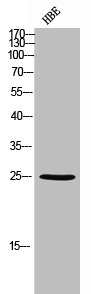
Western Blot analysis of HBE cells using Ebi3 Polyclonal Antibody
Western Blot analysis of HBE cells using Ebi3 Polyclonal Antibody
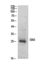
EBI3 Antibody
CSB-PA006315
ApplicationsWestern Blot, ELISA
Product group Antibodies
ReactivityHuman, Mouse
TargetEBI3
Overview
- SupplierCusabio
- Product NameEBI3 Antibody
- Delivery Days Customer20
- ApplicationsWestern Blot, ELISA
- CertificationResearch Use Only
- ClonalityPolyclonal
- ConjugateUnconjugated
- Gene ID10148
- Target nameEBI3
- Target descriptionEpstein-Barr virus induced 3
- Target synonymsIL-27B, IL27B, IL35B, interleukin-27 subunit beta, EBV-induced gene 3 protein, Epstein-Barr virus induced gene 3, IL-27 subunit beta, IL27 subunit, IL35 subunit, cytokine receptor, epstein-Barr virus-induced gene 3 protein
- HostRabbit
- IsotypeIgG
- Protein IDQ14213
- Protein NameInterleukin-27 subunit beta
- ReactivityHuman, Mouse
- Storage Instruction-20°C or -80°C
- UNSPSC41116161
Related products
Product group Antibodies
Anti-EBI3 AntibodyA99430
ApplicationsWestern Blot, ELISA
ReactivityHuman, Mouse
- SizePrice

Product group Antibodies
ApplicationsELISA, ELISpot Assay
ReactivityHuman
TargetEBI3
- SizePrice
Product group Antibodies
Anti-EBI3 (C-term) Antibody102-23783
ApplicationsWestern Blot
TargetEBI3
- SizePrice

Product group Antibodies
ApplicationsWestern Blot
ReactivityHuman, Mouse, Rat
TargetEBI3
- SizePrice
Product group Antibodies

EBI3 / IL-27B AntibodyLS-C831439
ApplicationsImmunoHistoChemistry
ReactivityHuman, Mouse, Rat
TargetEBI3
- SizePrice
Product group Antibodies
IL-27beta Recombinant AntibodyBSM-62257R
ApplicationsWestern Blot
ReactivityHuman, Mouse, Rat
TargetEBI3
- SizePrice

Product group Antibodies
Ebi3 Polyclonal AntibodyCAC08380
ApplicationsWestern Blot, ELISA, ImmunoHistoChemistry
TargetEBI3
- SizePrice
![Sandwich ELISA analysis of human IL-27 protein using GTX02984 IL27 antibody [MT27 + MT361] as coating antibody and GTX02919-02 EBI3 antibody [MT140] (Biotin) as detecting antibody.](https://www.genetex.com/upload/website/prouct_img/normal/GTX02919-02/GTX02919-02_20210507_ELISA_w_23053123_368.webp)
Product group Antibodies
EBI3 antibody [MT140] (Biotin)GTX02919-02
ApplicationsELISA
ReactivityHuman
TargetEBI3
- SizePrice