

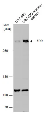
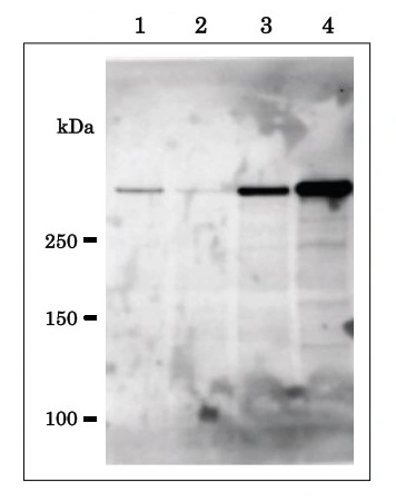
U87-MG whole cell and nuclear extracts (30 μg) were separated by 5% SDS-PAGE, and the membrane was blotted with EDD antibody (GTX130759) diluted at 1:500.
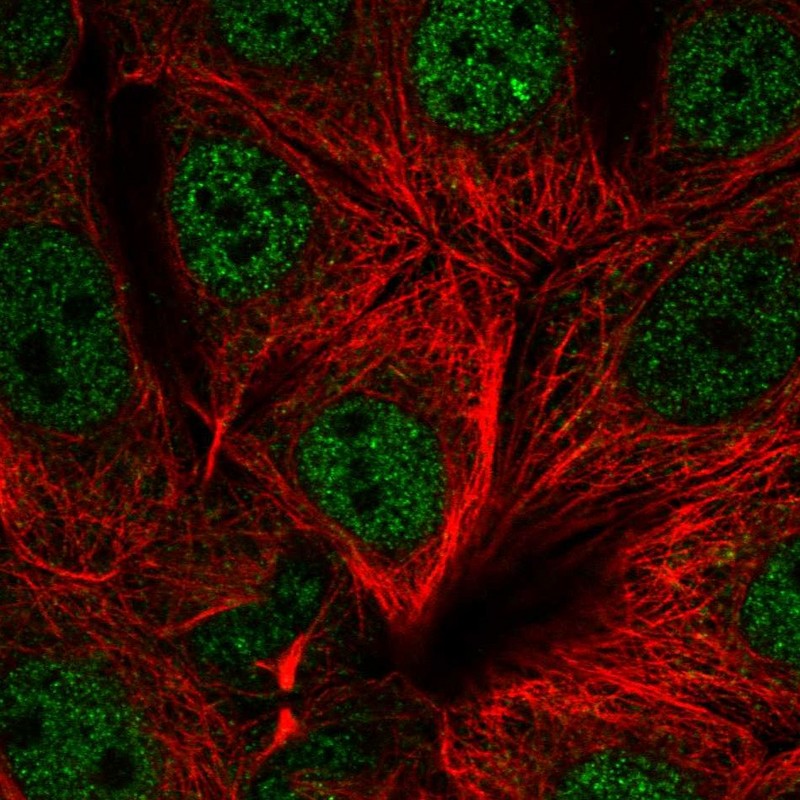
![EDD antibody detects EDD protein at nucleus by immunofluorescent analysis. Sample: U87-MG cells were fixed in 4% paraformaldehyde at RT for 15 min. Green: EDD protein stained by EDD antibody (GTX130759) diluted at 1:500. Red: alpha Tubulin, a cytoskeleton marker, stained by alpha Tubulin antibody [GT114] (GTX628802) diluted at 1:1000. Blue: Hoechst 33342 staining. Scale bar = 10 μm.](https://www.genetex.com/upload/website/prouct_img/normal/GTX130759/GTX130759_42431_20160602_IFA_w_23060523_481.webp "EDD antibody detects EDD protein at nucleus by immunofluorescent analysis. Sample: U87-MG cells were fixed in 4% paraformaldehyde at RT for 15 min. Green: EDD protein stained by EDD antibody (GTX130759) diluted at 1:500. Red: alpha Tubulin, a cytoskeleton marker, stained by alpha Tubulin antibody [GT114] (GTX628802) diluted at 1:1000. Blue: Hoechst 33342 staining. Scale bar = 10 μm.")
U87-MG whole cell and nuclear extracts (30 μg) were separated by 5% SDS-PAGE, and the membrane was blotted with EDD antibody (GTX130759) diluted at 1:500.
EDD antibody
GTX130759
ApplicationsImmunoFluorescence, Western Blot, ImmunoCytoChemistry
Product group Antibodies
ReactivityHuman
TargetUBR5
Overview
- SupplierGeneTex
- Product NameEDD antibody
- Delivery Days Customer9
- Application Supplier NoteWB: 1:500-1:3000. ICC/IF: 1:100-1:1000. *Optimal dilutions/concentrations should be determined by the researcher.Not tested in other applications.
- ApplicationsImmunoFluorescence, Western Blot, ImmunoCytoChemistry
- CertificationResearch Use Only
- ClonalityPolyclonal
- Concentration3.04 mg/ml
- ConjugateUnconjugated
- Gene ID51366
- Target nameUBR5
- Target descriptionubiquitin protein ligase E3 component n-recognin 5
- Target synonymsDD5, EDD, EDD1, HYD, E3 ubiquitin-protein ligase UBR5, E3 identified by differential display, E3 ubiquitin-protein ligase, HECT domain-containing 1, HECT-type E3 ubiquitin transferase UBR5, hyperplastic discs protein homolog, progestin-induced protein
- HostRabbit
- IsotypeIgG
- Protein IDO95071
- Protein NameE3 ubiquitin-protein ligase UBR5
- Scientific DescriptionThis gene encodes a progestin-induced protein, which belongs to the HECT (homology to E6-AP carboxyl terminus) family. The HECT family proteins function as E3 ubiquitin-protein ligases, targeting specific proteins for ubiquitin-mediated proteolysis. This gene is localized to chromosome 8q22 which is disrupted in a variety of cancers. This gene potentially has a role in regulation of cell proliferation or differentiation. [provided by RefSeq]
- ReactivityHuman
- Storage Instruction-20°C or -80°C,2°C to 8°C
- UNSPSC41116161
Datasheet
Related products
Product group Antibodies
UBR5 AntibodyCSB-PA004376
ApplicationsELISA, ImmunoHistoChemistry
ReactivityHuman, Mouse
TargetUBR5
- SizePrice
Product group Antibodies

UBR5 AntibodyLS-C748844
ApplicationsWestern Blot
ReactivityHuman
TargetUBR5
- SizePrice
Product group Antibodies
Anti-UBR5 AntibodyHPA053688
ApplicationsImmunoCytoChemistry
ReactivityHuman
TargetUBR5
- SizePrice
Product group Antibodies

Goat anti-EDD1 / HYDEB05920
ApplicationsELISA, ImmunoHistoChemistry
ReactivityBovine, Human, Mouse, Porcine, Rat
TargetUBR5
- SizePrice
Product group Antibodies
EDD antibody, C-termGTX89818
ApplicationsImmunoHistoChemistry, ImmunoHistoChemistry Paraffin
ReactivityHuman
TargetUBR5
- SizePrice
Product group Antibodies
UBR5 Polyclonal AntibodyBS-13052R
ApplicationsImmunoFluorescence, ELISA, ImmunoCytoChemistry, ImmunoHistoChemistry, ImmunoHistoChemistry Frozen, ImmunoHistoChemistry Paraffin
ReactivityCanine, Chicken, Equine, Human, Mouse, Porcine, Rat, Sheep
TargetUBR5
- SizePrice
Product group Antibodies
EDD antibodyGTX00902
ApplicationsImmunoFluorescence, Western Blot, ImmunoCytoChemistry
ReactivityHuman, Mouse
TargetUBR5
- SizePrice
Product group Antibodies
Anti-UBR5 Antibody144-60489
ApplicationsWestern Blot
ReactivityHuman
TargetUBR5
- SizePrice