

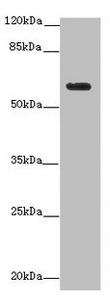
Western blot All lanes: EHD1 antibody at 12microg/ml + MDA-MB-231 whole cell lysate Secondary Goat polyclonal to rabbit IgG at 1/10000 dilution Predicted band size: 61 kDa Observed band size: 61 kDa

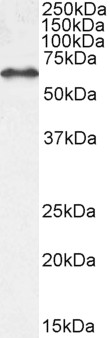
Western blot All lanes: EHD1 antibody at 12microg/ml + MDA-MB-231 whole cell lysate Secondary Goat polyclonal to rabbit IgG at 1/10000 dilution Predicted band size: 61 kDa Observed band size: 61 kDa
EHD1 Antibody
CSB-PA884470LA01HU
ApplicationsWestern Blot, ELISA, ImmunoHistoChemistry
Product group Antibodies
ReactivityHuman
TargetEHD1
Overview
- SupplierCusabio
- Product NameEHD1 Antibody
- Delivery Days Customer20
- ApplicationsWestern Blot, ELISA, ImmunoHistoChemistry
- CertificationResearch Use Only
- ClonalityPolyclonal
- ConjugateUnconjugated
- Gene ID10938
- Target nameEHD1
- Target descriptionEH domain containing 1
- Target synonymsH-PAST, HPAST1, PAST, PAST1, EH domain-containing protein 1, PAST homolog 1, testilin
- HostRabbit
- IsotypeIgG
- Protein IDQ9H4M9
- Protein NameEH domain-containing protein 1
- Scientific DescriptionATP- and membrane-binding protein that controls membrane reorganization/tubulation upon ATP hydrolysis. In vitro causes vesiculation of endocytic membranes (PubMed:24019528). Acts in early endocytic membrane fusion and membrane trafficking of recycling endosomes (PubMed:15020713, PubMed:17233914, PubMed:20801876). Recruited to endosomal membranes upon nerve growth factor stimulation, indirectly regulates neurite outgrowth (By similarity). Plays a role in myoblast fusion (By similarity). Involved in the unidirectional retrograde dendritic transport of endocytosed BACE1 and in efficient sorting of BACE1 to axons implicating a function in neuronal APP processing (By similarity). Plays a role in the formation of the ciliary vesicle (CV), an early step in cilium biogenesis. Proposed to be required for the fusion of distal appendage vesicles (DAVs) to form the CV by recruiting SNARE complex component SNAP29. Is required for recruitment of transition zone proteins CEP290, RPGRIP1L, TMEM67 and B9D2, and of IFT20 following DAV reorganization before Rab8-dependent ciliary membrane extension. Required for the loss of CCP110 form the mother centriole essential for the maturation of the basal body during ciliogenesis (PubMed:25686250).
- ReactivityHuman
- Storage Instruction-20°C or -80°C
- UNSPSC41116161
Related products
Product group Antibodies
Anti-EHD1 AntibodyA121141
ApplicationsFlow Cytometry, ImmunoFluorescence, Western Blot, ELISA, ImmunoHistoChemistry
ReactivityHuman, Mouse
- SizePrice

Product group Antibodies
Anti-EHD1 Antibody Picoband(r)A02168-1-CARRIER-FREE
ApplicationsFlow Cytometry, ImmunoFluorescence, Western Blot, ELISA, ImmunoCytoChemistry, ImmunoHistoChemistry, ImmunoHistoChemistry Frozen
ReactivityHuman, Mouse, Rat
TargetEHD1
- SizePrice
Product group Antibodies

Testilin / EHD1 AntibodyLS-C830029
ApplicationsWestern Blot, ELISA
ReactivityHuman, Mouse, Rat
TargetEHD1
- SizePrice
Product group Antibodies

Goat anti-EHD1EB06492
ApplicationsFlow Cytometry, ImmunoFluorescence, Western Blot, ELISA, ImmunoHistoChemistry
ReactivityBovine, Human, Mouse, Rat
TargetEHD1
- SizePrice

Product group Antibodies
Anti-EHD1 AntibodyHPA066751
ApplicationsImmunoCytoChemistry
ReactivityHuman
TargetEHD1
- SizePrice
Product group Antibodies
EHD1 Polyclonal AntibodyCAC14573
ApplicationsWestern Blot, ELISA, ImmunoHistoChemistry
TargetEHD1
- SizePrice
Product group Antibodies
EHD1 antibody, InternalGTX49037
ApplicationsWestern Blot, ImmunoHistoChemistry, ImmunoHistoChemistry Paraffin
ReactivityHuman
TargetEHD1
- SizePrice
Product group Antibodies
EHD1 Recombinant AntibodyBSM-62011R
ApplicationsFlow Cytometry, ImmunoFluorescence, ImmunoPrecipitation, Western Blot, ImmunoCytoChemistry, ImmunoHistoChemistry, ImmunoHistoChemistry Frozen, ImmunoHistoChemistry Paraffin
ReactivityHuman, Mouse, Rat
TargetEHD1
- SizePrice