

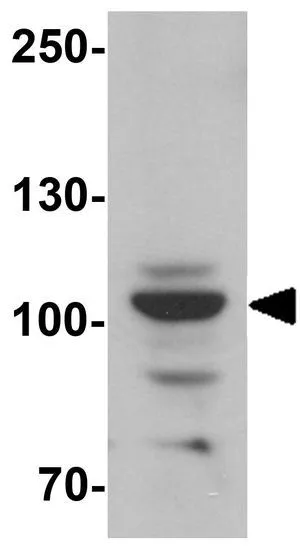
WB analysis of various samples using GTX03246 Epac1 antibody [GT1334]. Dilution : 1:1000 Loading : 25μg per lane
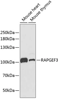
![Various whole cell extracts (30 μg) were separated by 10% SDS-PAGE, and the membrane was blotted with Epac1 antibody [GT1334] (GTX03246) diluted at 1:1000. The HRP-conjugated anti-rabbit IgG antibody (GTX213110-01) was used to detect the primary antibody, and the signal was developed with Trident ECL plus-Enhanced.](https://www.genetex.com/upload/website/prouct_img/normal/GTX03246/GTX03246_4000000908_20210709_WB_w_23053123_813.webp "Various whole cell extracts (30 μg) were separated by 10% SDS-PAGE, and the membrane was blotted with Epac1 antibody [GT1334] (GTX03246) diluted at 1:1000. The HRP-conjugated anti-rabbit IgG antibody (GTX213110-01) was used to detect the primary antibody, and the signal was developed with Trident ECL plus-Enhanced.")
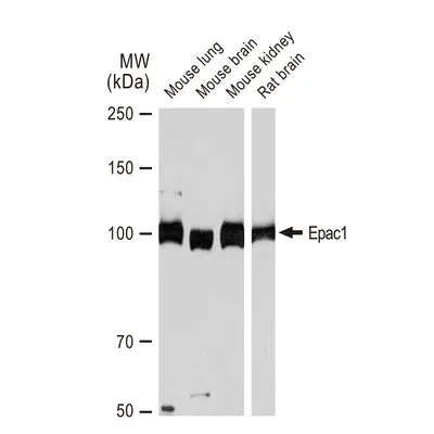
![Various tissue extracts (50 μg) were separated by 10% SDS-PAGE, and the membrane was blotted with Epac1 antibody [GT1334] (GTX03246) diluted at 1:1000. The HRP-conjugated anti-rabbit IgG antibody (GTX213110-01) was used to detect the primary antibody, and the signal was developed with Trident ECL plus-Enhanced.](https://www.genetex.com/upload/website/prouct_img/normal/GTX03246/GTX03246_4000000908_20210709_WB_M_R_w_23053123_712.webp "Various tissue extracts (50 μg) were separated by 10% SDS-PAGE, and the membrane was blotted with Epac1 antibody [GT1334] (GTX03246) diluted at 1:1000. The HRP-conjugated anti-rabbit IgG antibody (GTX213110-01) was used to detect the primary antibody, and the signal was developed with Trident ECL plus-Enhanced.")
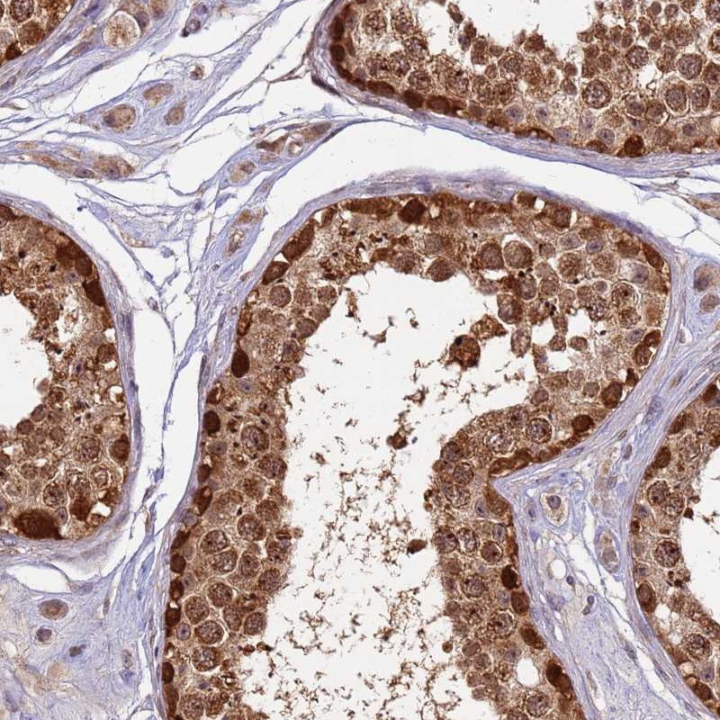
![IHC-P analysis of human placenta tissue section using GTX03246 Epac1 antibody [GT1334]. Dilution : 1:100](https://www.genetex.com/upload/website/prouct_img/normal/GTX03246/GTX03246_20210615_IHC-P_23_w_23053123_598.webp "IHC-P analysis of human placenta tissue section using GTX03246 Epac1 antibody [GT1334]. Dilution : 1:100")
WB analysis of various samples using GTX03246 Epac1 antibody [GT1334]. Dilution : 1:1000 Loading : 25μg per lane
Epac1 antibody [GT1334]
GTX03246
ApplicationsWestern Blot, ImmunoHistoChemistry, ImmunoHistoChemistry Paraffin
Product group Antibodies
ReactivityHuman, Mouse, Rat
TargetRAPGEF3
Overview
- SupplierGeneTex
- Product NameEpac1 antibody [GT1334]
- Delivery Days Customer9
- Application Supplier NoteWB: 1:500 - 1:2000. IHC-P: 1:50 - 1:200. *Optimal dilutions/concentrations should be determined by the researcher.Not tested in other applications.
- ApplicationsWestern Blot, ImmunoHistoChemistry, ImmunoHistoChemistry Paraffin
- CertificationResearch Use Only
- ClonalityMonoclonal
- Clone IDGT1334
- Concentration0.5 mg/ml
- ConjugateUnconjugated
- Gene ID10411
- Target nameRAPGEF3
- Target descriptionRap guanine nucleotide exchange factor 3
- Target synonymsCAMP-GEFI, EPAC, EPAC1, HSU79275, bcm910, rap guanine nucleotide exchange factor 3, 9330170P05Rik, EPAC 1, Rap guanine nucleotide exchange factor (GEF) 3, Rap1 guanine-nucleotide-exchange factor directly activated by cAMP, cAMP-regulated guanine nucleotide exchange factor I, exchange factor directly activated by cAMP 1, exchange protein directly activated by cAMP 1
- HostRabbit
- IsotypeIgG
- Protein IDO95398
- Protein NameRap guanine nucleotide exchange factor 3
- ReactivityHuman, Mouse, Rat
- Storage Instruction-20°C or -80°C,2°C to 8°C
- UNSPSC41116161
Datasheet
Related products
Product group Antibodies
Anti-RAPGEF3 AntibodyA96548
ApplicationsWestern Blot, ELISA
ReactivityHuman, Mouse, Rat
- SizePrice

Product group Antibodies
Anti-Epac1/RAPGEF3 Antibody Picoband(r)A02483-2-CARRIER-FREE
ApplicationsFlow Cytometry, Western Blot, ELISA
ReactivityHuman, Monkey, Mouse, Rat
TargetRAPGEF3
- SizePrice
Product group Antibodies
Anti-RAPGEF3 Antibody144-02199
ApplicationsImmunoFluorescence, Western Blot
ReactivityHuman, Mouse
TargetRAPGEF3
- SizePrice
Product group Antibodies
Epac1 Recombinant Antibody, AbBy Fluor-350 ConjugatedBSM-61700R-BF350
ApplicationsImmunoFluorescence, Western Blot
ReactivityHuman, Mouse, Rat
TargetRAPGEF3
- SizePrice
Product group Antibodies
RAPGEF3 AntibodyCSB-PA006508
ApplicationsWestern Blot, ELISA
ReactivityHuman, Mouse, Rat
TargetRAPGEF3
- SizePrice
Product group Antibodies

ApplicationsWestern Blot, ELISA
ReactivityHuman, Mouse, Rat
TargetRAPGEF3
- SizePrice
Product group Antibodies
Anti-RAPGEF3 AntibodyHPA040365
ApplicationsImmunoHistoChemistry
ReactivityHuman
TargetRAPGEF3
- SizePrice
Product group Antibodies
Epac1 antibodyGTX54723
ApplicationsImmunoFluorescence, Western Blot, ImmunoCytoChemistry
ReactivityHuman, Mouse
TargetRAPGEF3
- SizePrice
Product group Antibodies
Epac1 antibodyGTX32047
ApplicationsWestern Blot, ELISA, ImmunoHistoChemistry, ImmunoHistoChemistry Paraffin
ReactivityHuman, Mouse, Rat
TargetRAPGEF3
- SizePrice

Product group Antibodies
References
Epac1 antibodyGTX41235
ApplicationsImmunoFluorescence, Western Blot, ELISA, ImmunoCytoChemistry, ImmunoHistoChemistry
ReactivityEquine, Human, Mouse, Rat
TargetRAPGEF3
- SizePrice