

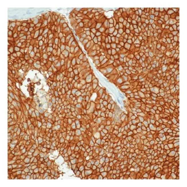
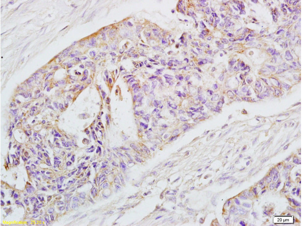
IHC-P analysis of human breast carcinoma tissue using GTX28667 EpCAM antibody [VU-1D9].
IHC-P analysis of human breast carcinoma tissue using GTX28667 EpCAM antibody [VU-1D9].
EpCAM antibody [VU-1D9]
GTX28667
ApplicationsFlow Cytometry, ImmunoFluorescence, Western Blot, ImmunoCytoChemistry, ImmunoHistoChemistry, ImmunoHistoChemistry Paraffin
Product group Antibodies
ReactivityHuman
TargetEPCAM
Overview
- SupplierGeneTex
- Product NameEpCAM antibody [VU-1D9]
- Delivery Days Customer9
- Application Supplier NoteIHC-P: 1-2 microg/ml. *Optimal dilutions/concentrations should be determined by the researcher.Not tested in other applications.
- ApplicationsFlow Cytometry, ImmunoFluorescence, Western Blot, ImmunoCytoChemistry, ImmunoHistoChemistry, ImmunoHistoChemistry Paraffin
- CertificationResearch Use Only
- ClonalityMonoclonal
- Clone IDVU-1D9
- Concentration200 ug/ml
- ConjugateUnconjugated
- Gene ID4072
- Target nameEPCAM
- Target descriptionepithelial cell adhesion molecule
- Target synonymsBer-Ep4, BerEp4, DIAR5, EGP-2, EGP314, EGP40, ESA, HNPCC8, KS1/4, KSA, LYNCH8, M4S1, MIC18, MK-1, MOC-31, TACSTD1, TROP1, epithelial cell adhesion molecule, adenocarcinoma-associated antigen, cell surface glycoprotein Trop-1, epithelial glycoprotein 314, human epithelial glycoprotein-2, major gastrointestinal tumor-associated protein GA733-2, membrane component, chromosome 4, surface marker (35kD glycoprotein), trophoblast cell surface antigen 1, tumor-associated calcium signal transducer 1
- HostMouse
- IsotypeIgG1
- Protein IDP16422
- Protein NameEpithelial cell adhesion molecule
- Scientific DescriptionThis gene encodes a carcinoma-associated antigen and is a member of a family that includes at least two type I membrane proteins. This antigen is expressed on most normal epithelial cells and gastrointestinal carcinomas and functions as a homotypic calcium-independent cell adhesion molecule. The antigen is being used as a target for immunotherapy treatment of human carcinomas. Mutations in this gene result in congenital tufting enteropathy. [provided by RefSeq, Dec 2008]
- ReactivityHuman
- Storage Instruction2°C to 8°C
- UNSPSC12352203
References
- Twigger AJ, Engelbrecht LK, Bach K, et al. Transcriptional changes in the mammary gland during lactation revealed by single cell sequencing of cells from human milk. Nat Commun. 2022,13(1):562. doi: 10.1038/s41467-021-27895-0Read this paper
- Mikami H, Kawaguchi M, Huang CJ, et al. Virtual-freezing fluorescence imaging flow cytometry. Nat Commun. 2020,11(1):1162. doi: 10.1038/s41467-020-14929-2Read this paper
- Yoshida M, Hibino K, Yamamoto S, et al. Preferential capture of EpCAM-expressing extracellular vesicles on solid surfaces coated with an aptamer-conjugated zwitterionic polymer. Biotechnol Bioeng. 2018,115(3):536-544. doi: 10.1002/bit.26489Read this paper
- Kershaw S, Cummings J, Morris K, et al. Optimisation of immunofluorescence methods to determine MCT1 and MCT4 expression in circulating tumour cells. BMC Cancer. 2015,15:387. doi: 10.1186/s12885-015-1382-yRead this paper
- Lyberopoulou A, Aravantinos G, Efstathopoulos EP, et al. Mutational analysis of circulating tumor cells from colorectal cancer patients and correlation with primary tumor tissue. PLoS One. 2015,10(4):e0123902. doi: 10.1371/journal.pone.0123902Read this paper
- Ma R, Fredriksson I, Karthik GM, et al. Superficial scrapings from breast tumors is a source for biobanking and research purposes. Lab Invest. 2014,94(7):796-805. doi: 10.1038/labinvest.2014.65Read this paper
- Chen HH, Lin MW, Tien WT, et al. High-purity separation of cancer cells by optically induced dielectrophoresis. J Biomed Opt. 2014,19(4):045002. doi: 10.1117/1.JBO.19.4.045002Read this paper
- Okuda H, Kobayashi A, Xia B, et al. Hyaluronan synthase HAS2 promotes tumor progression in bone by stimulating the interaction of breast cancer stem-like cells with macrophages and stromal cells. Cancer Res. 2012,72(2):537-47. doi: 10.1158/0008-5472.CAN-11-1678Read this paper
- Kimura O, Takahashi T, Ishii N, et al. Characterization of the epithelial cell adhesion molecule (EpCAM)+ cell population in hepatocellular carcinoma cell lines. Cancer Sci. 2010,101(10):2145-55. doi: 10.1111/j.1349-7006.2010.01661.xRead this paper
Datasheet
Related products
Product group Antibodies
Epcam Polyclonal AntibodyCAC11319
ApplicationsImmunoFluorescence, Western Blot, ELISA, ImmunoHistoChemistry
ReactivityRat
TargetEPCAM
- SizePrice
Product group Antibodies
References
EpCAM Polyclonal AntibodyBS-1513R
ApplicationsFlow Cytometry, ImmunoFluorescence, Western Blot, ELISA, ImmunoCytoChemistry, ImmunoHistoChemistry, ImmunoHistoChemistry Frozen, ImmunoHistoChemistry Paraffin
ReactivityHuman, Mouse, Rat
TargetEPCAM
- SizePrice
Product group Antibodies
Anti-EpCAM AntibodyA85204
ApplicationsWestern Blot, ELISA
ReactivityHuman
- SizePrice
Product group Antibodies
Anti-EPCAM Antibody144-01177
ApplicationsWestern Blot, ImmunoHistoChemistry
ReactivityHuman, Mouse
TargetEPCAM
- SizePrice

Product group Antibodies
Anti-EPCAM AntibodyAMAB91411
ApplicationsWestern Blot, ImmunoHistoChemistry
ReactivityHuman
TargetEPCAM
- SizePrice
Product group Antibodies

Anti-EpCAM [AUA1]Ab00609-1.1
ApplicationsFlow Cytometry, ImmunoFluorescence, ELISA, ImmunoHistoChemistry
ReactivityHuman
TargetEPCAM
- SizePrice
Product group Antibodies

ApplicationsWestern Blot, ELISA
ReactivityHuman
TargetEPCAM
- SizePrice
![FACS analysis of MCF-7 cells using GTX00606-07 EpCAM antibody [VU-1D9] (APC).](https://www.genetex.com/upload/website/prouct_img/normal/GTX00606-07/GTX00606-07_20191025_AP_006_105_w_23053121_233.webp)
Product group Antibodies
References
EpCAM antibody [VU-1D9] (APC)GTX00606-07
ApplicationsFlow Cytometry
ReactivityHuman
TargetEPCAM
- SizePrice