

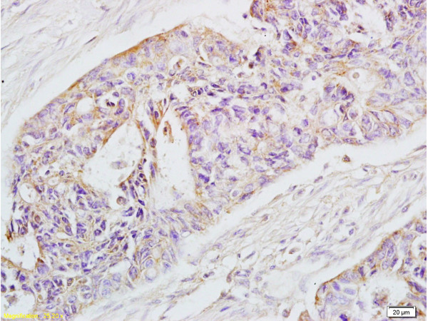
Formalin-fixed and paraffin-embedded human rectal carcinoma labeled with Rabbit Anti-EpCAM Polyclonal Antibody (bs-1513R), Unconjugated 1:200 followed by conjugation to the secondary antibody and DAB staining
. The cells were then incubated in 1 X PBS containing 0.5% BSA + 10% goat serum to block non-specific protein-protein interactions for 15 min at room temperature. Staining with EpCAM Polyclonal Antibody, Unconjugated (bs-1513R) was performed at 1:100 for 30 min on ice. The secondary antibody, Goat anti-rabbit IgG-PE, was used at a 1:200 dilution for 30 minutes on ice. Primary antibody staining (green) is compared to compared to unstained cells (dark blue), secondary only (light blue), and isotype control (orange).")
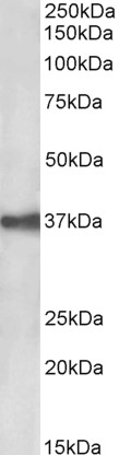
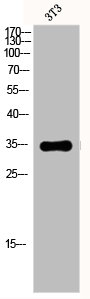
 at 1:1000 dilution and 4˚C overnight incubation. Followed by conjugated secondary antibody incubation at 1:20000 for 60 min at 37˚C.")
 at 1:1000 dilution and 4˚C overnight incubation. Followed by conjugated secondary antibody incubation at 1:20000 for 60 min at 37˚C.")
 for 15min; Block endogenous peroxidase by 3% hydrogen peroxide for 20 minutes; Blocking buffer (normal goat serum) at 37°C for 30min; Antibody incubation with EpCAM/CD326 Polyclonal Antibody, Unconjugated (bs-1513R) at 1:400 overnight at 4°C, DAB staining.")
 at 1:1000 dilution and 4˚C overnight incubation. Followed by conjugated secondary antibody incubation at 1:20000 for 60 min at 37˚C.")
 at 1:1000 dilution and 4˚C overnight incubation. Followed by conjugated secondary antibody incubation at 1:20000 for 60 min at 37˚C.")
at 1:100 dilution in blocking buffer and incubated for 30 min at room temperature, washed twice with 2%BSA in PBS, followed by secondary antibody incubation for 40 min at room temperature. Acquisitions of 20,000 events were performed. Cells stained with primary antibody (green), and isotype control (orange).")
at 1:50 dilution in blocking buffer and incubated for 30 min at room temperature, washed twice with 2%BSA in PBS, followed by secondary antibody incubation for 40 min at room temperature. Acquisitions of 20,000 events were performed. Cells stained with primary antibody (green), and isotype control (orange).")
; Antigen retrieval by boiling in sodium citrate buffer (pH6.0) for 15min; Block endogenous peroxidase by 3% hydrogen peroxide for 20 minutes; Blocking buffer (normal goat serum) at 37°C for 30min; Antibody incubation with (EpCAM) Polyclonal Antibody, Unconjugated (bs-1513R) at 1:200 overnight at 4°C, followed by operating according to SP Kit(Rabbit) (sp-0023) instructionsand DAB staining.")
Formalin-fixed and paraffin-embedded human rectal carcinoma labeled with Rabbit Anti-EpCAM Polyclonal Antibody (bs-1513R), Unconjugated 1:200 followed by conjugation to the secondary antibody and DAB staining
EpCAM Polyclonal Antibody
BS-1513R
ApplicationsFlow Cytometry, ImmunoFluorescence, Western Blot, ELISA, ImmunoCytoChemistry, ImmunoHistoChemistry, ImmunoHistoChemistry Frozen, ImmunoHistoChemistry Paraffin
Product group Antibodies
ReactivityHuman, Mouse, Rat
TargetEPCAM
Overview
- SupplierBioss
- Product NameEpCAM Polyclonal Antibody
- Delivery Days Customer16
- ApplicationsFlow Cytometry, ImmunoFluorescence, Western Blot, ELISA, ImmunoCytoChemistry, ImmunoHistoChemistry, ImmunoHistoChemistry Frozen, ImmunoHistoChemistry Paraffin
- Applications SupplierWB(1:300-5000), ELISA(1:500-1000), FCM(1:20-100), IHC-P(1:200-400), IHC-F(1:100-500), IF(IHC-P)(1:50-200), IF(IHC-F)(1:50-200), IF(ICC)(1:50-200)
- CertificationResearch Use Only
- ClonalityPolyclonal
- Concentration1 ug/ul
- ConjugateUnconjugated
- Gene ID4072
- Target nameEPCAM
- Target descriptionepithelial cell adhesion molecule
- Target synonymsBer-Ep4, BerEp4, DIAR5, EGP-2, EGP314, EGP40, ESA, HNPCC8, KS1/4, KSA, LYNCH8, M4S1, MIC18, MK-1, MOC-31, TACSTD1, TROP1, epithelial cell adhesion molecule, adenocarcinoma-associated antigen, cell surface glycoprotein Trop-1, epithelial glycoprotein 314, human epithelial glycoprotein-2, major gastrointestinal tumor-associated protein GA733-2, membrane component, chromosome 4, surface marker (35kD glycoprotein), trophoblast cell surface antigen 1, tumor-associated calcium signal transducer 1
- HostRabbit
- IsotypeIgG
- Protein IDP16422
- Protein NameEpithelial cell adhesion molecule
- ReactivityHuman, Mouse, Rat
- Storage Instruction-20°C
- UNSPSC41116161
References
- Epithelial cell adhesion molecule fragments and signaling in primary human liver cells. Gerlach JC et al., 2018 Jun, J Cell PhysiolRead this paper
Datasheet
Related products
Product group Antibodies
Anti-EpCAM AntibodyA85204
ApplicationsWestern Blot, ELISA
ReactivityHuman
- SizePrice
Product group Antibodies

Anti-EpCAM [AUA1]Ab00609-1.1
ApplicationsFlow Cytometry, ImmunoFluorescence, ELISA, ImmunoHistoChemistry
ReactivityHuman
TargetEPCAM
- SizePrice
Product group Antibodies
Anti-EPCAM Antibody144-01177
ApplicationsWestern Blot, ImmunoHistoChemistry
ReactivityHuman, Mouse
TargetEPCAM
- SizePrice

Product group Antibodies
Anti-EPCAM AntibodyAMAB91411
ApplicationsWestern Blot, ImmunoHistoChemistry
ReactivityHuman
TargetEPCAM
- SizePrice
Product group Antibodies

EPCAM Antibody (clone G8.8, PE)LS-C810981
ApplicationsFlow Cytometry
ReactivityMouse
TargetEPCAM
- SizePrice
Product group Antibodies
EPCAM AntibodyCSB-PA002357
ApplicationsImmunoFluorescence, Western Blot, ELISA
ReactivityHuman
TargetEPCAM
- SizePrice
Product group Antibodies

ApplicationsWestern Blot, ELISA
ReactivityHuman
TargetEPCAM
- SizePrice
Product group Antibodies
Epcam Polyclonal AntibodyCAC11319
ApplicationsImmunoFluorescence, Western Blot, ELISA, ImmunoHistoChemistry
ReactivityRat
TargetEPCAM
- SizePrice
Product group Antibodies

ApplicationsImmunoHistoChemistry, ImmunoHistoChemistry Frozen, ImmunoHistoChemistry Paraffin
ReactivityHuman
TargetEPCAM
- SizePrice