

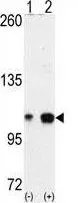
WB analysis of 293 cell lysate (2 ug/lane) either nontransfected (Lane 1) or transiently transfected with the EPHA4 (Lane 2) using GTX25396 EphA4 antibody.


WB analysis of 293 cell lysate (2 ug/lane) either nontransfected (Lane 1) or transiently transfected with the EPHA4 (Lane 2) using GTX25396 EphA4 antibody.
EphA4 antibody
GTX25396
ApplicationsWestern Blot, ImmunoHistoChemistry, ImmunoHistoChemistry Paraffin
Product group Antibodies
ReactivityHuman, Mouse
TargetEPHA4
Overview
- SupplierGeneTex
- Product NameEphA4 antibody
- Delivery Days Customer7
- Application Supplier NoteWB: 1:1000. IHC-P: 1:50-1:100. *Optimal dilutions/concentrations should be determined by the researcher.Not tested in other applications.
- ApplicationsWestern Blot, ImmunoHistoChemistry, ImmunoHistoChemistry Paraffin
- CertificationResearch Use Only
- ClonalityPolyclonal
- ConjugateUnconjugated
- Gene ID2043
- Target nameEPHA4
- Target descriptionEPH receptor A4
- Target synonymsEK8, HEK8, SEK, TYRO1, ephrin type-A receptor 4, EPH-like kinase 8, TYRO1 protein tyrosine kinase, receptor protein-tyrosine kinase HEK8, tyrosine-protein kinase TYRO1, tyrosine-protein kinase receptor SEK
- HostRabbit
- IsotypeIgG
- Protein IDP54764
- Protein NameEphrin type-A receptor 4
- Scientific DescriptionThis gene belongs to the ephrin receptor subfamily of the protein-tyrosine kinase family. EPH and EPH-related receptors have been implicated in mediating developmental events, particularly in the nervous system. Receptors in the EPH subfamily typically have a single kinase domain and an extracellular region containing a Cys-rich domain and 2 fibronectin type III repeats. The ephrin receptors are divided into 2 groups based on the similarity of their extracellular domain sequences and their affinities for binding ephrin-A and ephrin-B ligands. Multiple transcript variants encoding different isoforms have been found for this gene. [provided by RefSeq, Jan 2015]
- ReactivityHuman, Mouse
- Storage Instruction-20°C or -80°C,2°C to 8°C
- UNSPSC41116161
Datasheet
Related products
Product group Antibodies
Anti-EPHA4 AntibodyA46071
ApplicationsImmunoHistoChemistry
ReactivityHuman, Mouse
- SizePrice
Product group Antibodies

EPHA4 (Phospho-Tyr596) AntibodyABX012736
ApplicationsWestern Blot, ELISA
- SizePrice
Product group Antibodies
Anti-EPHA4 Antibody144-08346
ApplicationsWestern Blot
ReactivityHuman, Mouse, Rat
TargetEPHA4
- SizePrice
Product group Antibodies
ApplicationsImmunoFluorescence, ELISA, ImmunoCytoChemistry, ImmunoHistoChemistry, ImmunoHistoChemistry Frozen, ImmunoHistoChemistry Paraffin
ReactivityCanine, Chicken, Equine, Human, Mouse, Porcine, Rabbit, Rat
TargetEPHA4
- SizePrice
Product group Antibodies
EPHA4 AntibodyCSB-PA007724LA01HU
ApplicationsImmunoFluorescence, ELISA, ImmunoHistoChemistry
ReactivityHuman
TargetEPHA4
- SizePrice
Product group Antibodies
EPHA4 Polyclonal AntibodyCAC12899
ApplicationsImmunoFluorescence, ELISA, ImmunoHistoChemistry
TargetEPHA4
- SizePrice
Product group Antibodies

ApplicationsWestern Blot
ReactivityChicken, Human, Mouse
TargetEPHA4
- SizePrice
![WB analysis of truncated Trx-EphA4 recombinant protein (1) andtruncated GST-EphA4(aa777-986) recombinant protein (2) using GTX83018 EphA4 antibody [7D3D4].](https://www.genetex.com/upload/website/prouct_img/normal/GTX83018/GTX83018_20170912_WB_w_23061322_402.webp)
Product group Antibodies
EphA4 antibody [7D3D4]GTX83018
ApplicationsWestern Blot, ELISA
ReactivityHuman
TargetEPHA4
- SizePrice
![Non-transfected (–) and transfected (+) 293T whole cell extracts (30 μg) were separated by 7.5% SDS-PAGE, and the membrane was blotted with EphA4 antibody [N3C2], Internal (GTX104109) diluted at 1:5000. The HRP-conjugated anti-rabbit IgG antibody (GTX213110-01) was used to detect the primary antibody.](https://www.genetex.com/upload/website/prouct_img/normal/GTX104109/GTX104109_40583_20180727_WB_B_w_23060120_399.webp)
Product group Antibodies
EphA4 antibody [N3C2], InternalGTX104109
ApplicationsWestern Blot
ReactivityHuman, Mouse, Rat
TargetEPHA4
- SizePrice
![Non-transfected (–) and transfected (+) 293T whole cell extracts (30 μg) were separated by 7.5% SDS-PAGE, and the membrane was blotted with EphA4 antibody [N3C2-2], Internal (GTX111602) diluted at 1:5000. The HRP-conjugated anti-rabbit IgG antibody (GTX213110-01) was used to detect the primary antibody.](https://www.genetex.com/upload/website/prouct_img/normal/GTX111602/GTX111602_40618_20180727_WB_B_w_23060500_777.webp)
Product group Antibodies
EphA4 antibody [N3C2-2], InternalGTX111602
ApplicationsWestern Blot, ImmunoHistoChemistry, ImmunoHistoChemistry Paraffin
ReactivityHuman, Mouse, Rat
TargetEPHA4
- SizePrice