

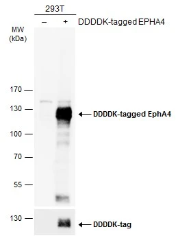
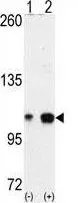
Non-transfected (–) and transfected (+) 293T whole cell extracts (30 μg) were separated by 7.5% SDS-PAGE, and the membrane was blotted with EphA4 antibody [N3C2-2], Internal (GTX111602) diluted at 1:5000. The HRP-conjugated anti-rabbit IgG antibody (GTX213110-01) was used to detect the primary antibody.
 A: HeLa 7.5% SDS PAGE GTX111602 diluted at 1:1000")
 were separated by 7.5% SDS-PAGE, and the membrane was blotted with EphA4 antibody (GTX111602) diluted at 1:1000. The HRP-conjugated anti-rabbit IgG antibody (GTX213110-01) was used to detect the primary antibody.")
![Mouse tissue extract (50 μg) was separated by 7.5% SDS-PAGE, and the membrane was blotted with EphA4 antibody [N3C2], Internal (GTX111602) diluted at 1:1000. The HRP-conjugated anti-rabbit IgG antibody (GTX213110-01) was used to detect the primary antibody.](https://www.genetex.com/upload/website/prouct_img/normal/GTX111602/GTX111602_40618_20170608_WB_M_hippocampus_w_23060500_667.webp "Mouse tissue extract (50 μg) was separated by 7.5% SDS-PAGE, and the membrane was blotted with EphA4 antibody [N3C2], Internal (GTX111602) diluted at 1:1000. The HRP-conjugated anti-rabbit IgG antibody (GTX213110-01) was used to detect the primary antibody.")
![Eph receptor A4 antibody [N3C2-2], Internal detects Eph receptor A4 protein by western blot analysis. Whole cell extracts (30 μg) was separated by 5 % SDS-PAGE, and blotted with Eph receptor A4 antibody [N3C2-2], Internal (GTX111602) diluted by 1:1000](https://www.genetex.com/upload/website/prouct_img/normal/GTX111602/GTX111602_40618_WB_2_w_23060500_742.webp "Eph receptor A4 antibody [N3C2-2], Internal detects Eph receptor A4 protein by western blot analysis. Whole cell extracts (30 μg) was separated by 5 % SDS-PAGE, and blotted with Eph receptor A4 antibody [N3C2-2], Internal (GTX111602) diluted by 1:1000")
Non-transfected (–) and transfected (+) 293T whole cell extracts (30 μg) were separated by 7.5% SDS-PAGE, and the membrane was blotted with EphA4 antibody [N3C2-2], Internal (GTX111602) diluted at 1:5000. The HRP-conjugated anti-rabbit IgG antibody (GTX213110-01) was used to detect the primary antibody.
EphA4 antibody [N3C2-2], Internal
GTX111602
ApplicationsWestern Blot, ImmunoHistoChemistry, ImmunoHistoChemistry Paraffin
Product group Antibodies
ReactivityHuman, Mouse, Rat
TargetEPHA4
Overview
- SupplierGeneTex
- Product NameEphA4 antibody [N3C2-2], Internal
- Delivery Days Customer9
- Application Supplier NoteWB: 1:1000-1:10000. *Optimal dilutions/concentrations should be determined by the researcher.Not tested in other applications.
- ApplicationsWestern Blot, ImmunoHistoChemistry, ImmunoHistoChemistry Paraffin
- CertificationResearch Use Only
- ClonalityPolyclonal
- Concentration0.97 mg/ml
- ConjugateUnconjugated
- Gene ID2043
- Target nameEPHA4
- Target descriptionEPH receptor A4
- Target synonymsEK8, HEK8, SEK, TYRO1, ephrin type-A receptor 4, EPH-like kinase 8, TYRO1 protein tyrosine kinase, receptor protein-tyrosine kinase HEK8, tyrosine-protein kinase TYRO1, tyrosine-protein kinase receptor SEK
- HostRabbit
- IsotypeIgG
- Protein IDP54764
- Protein NameEphrin type-A receptor 4
- Scientific DescriptionThis gene belongs to the ephrin receptor subfamily of the protein-tyrosine kinase family. EPH and EPH-related receptors have been implicated in mediating developmental events, particularly in the nervous system. Receptors in the EPH subfamily typically have a single kinase domain and an extracellular region containing a Cys-rich domain and 2 fibronectin type III repeats. The ephrin receptors are divided into 2 groups based on the similarity of their extracellular domain sequences and their affinities for binding ephrin-A and ephrin-B ligands. [provided by RefSeq]
- ReactivityHuman, Mouse, Rat
- Storage Instruction-20°C or -80°C,2°C to 8°C
- UNSPSC41116161
Datasheet
Related products
Product group Antibodies
Anti-EPHA4 AntibodyA46071
ApplicationsImmunoHistoChemistry
ReactivityHuman, Mouse
- SizePrice
Product group Antibodies

EPHA4 (Phospho-Tyr596) AntibodyABX012736
ApplicationsWestern Blot, ELISA
- SizePrice
Product group Antibodies
Anti-EPHA4 Antibody144-08346
ApplicationsWestern Blot
ReactivityHuman, Mouse, Rat
TargetEPHA4
- SizePrice
Product group Antibodies
ApplicationsImmunoFluorescence, ELISA, ImmunoCytoChemistry, ImmunoHistoChemistry, ImmunoHistoChemistry Frozen, ImmunoHistoChemistry Paraffin
ReactivityCanine, Chicken, Equine, Human, Mouse, Porcine, Rabbit, Rat
TargetEPHA4
- SizePrice
Product group Antibodies
EPHA4 AntibodyCSB-PA007724LA01HU
ApplicationsImmunoFluorescence, ELISA, ImmunoHistoChemistry
ReactivityHuman
TargetEPHA4
- SizePrice
Product group Antibodies
EPHA4 Polyclonal AntibodyCAC12899
ApplicationsImmunoFluorescence, ELISA, ImmunoHistoChemistry
TargetEPHA4
- SizePrice
Product group Antibodies
EphA4 antibodyGTX25396
ApplicationsWestern Blot, ImmunoHistoChemistry, ImmunoHistoChemistry Paraffin
ReactivityHuman, Mouse
TargetEPHA4
- SizePrice
Product group Antibodies

ApplicationsWestern Blot
ReactivityChicken, Human, Mouse
TargetEPHA4
- SizePrice
![WB analysis of truncated Trx-EphA4 recombinant protein (1) andtruncated GST-EphA4(aa777-986) recombinant protein (2) using GTX83018 EphA4 antibody [7D3D4].](https://www.genetex.com/upload/website/prouct_img/normal/GTX83018/GTX83018_20170912_WB_w_23061322_402.webp)
Product group Antibodies
EphA4 antibody [7D3D4]GTX83018
ApplicationsWestern Blot, ELISA
ReactivityHuman
TargetEPHA4
- SizePrice
![Non-transfected (–) and transfected (+) 293T whole cell extracts (30 μg) were separated by 7.5% SDS-PAGE, and the membrane was blotted with EphA4 antibody [N3C2], Internal (GTX104109) diluted at 1:5000. The HRP-conjugated anti-rabbit IgG antibody (GTX213110-01) was used to detect the primary antibody.](https://www.genetex.com/upload/website/prouct_img/normal/GTX104109/GTX104109_40583_20180727_WB_B_w_23060120_399.webp)
Product group Antibodies
EphA4 antibody [N3C2], InternalGTX104109
ApplicationsWestern Blot
ReactivityHuman, Mouse, Rat
TargetEPHA4
- SizePrice