

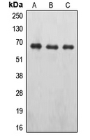
WB analysis of A498 (A), pervanadate-treated MCF7 (B), NIH3T3 (C) whole cell lysates using GTX32177 EPO receptor (phospho Tyr426) antibody.
WB analysis of A498 (A), pervanadate-treated MCF7 (B), NIH3T3 (C) whole cell lysates using GTX32177 EPO receptor (phospho Tyr426) antibody.
EPO receptor (phospho Tyr426) antibody
GTX32177
ApplicationsWestern Blot
Product group Antibodies
ReactivityHuman, Mouse
TargetEPOR
Overview
- SupplierGeneTex
- Product NameEPO receptor (phospho Tyr426) antibody
- Delivery Days Customer9
- Application Supplier NoteWB: 1:500 - 1:1000. *Optimal dilutions/concentrations should be determined by the researcher.Not tested in other applications.
- ApplicationsWestern Blot
- CertificationResearch Use Only
- ClonalityPolyclonal
- ConjugateUnconjugated
- Gene ID2057
- Target nameEPOR
- Target descriptionerythropoietin receptor
- Target synonymsEPO-R, erythropoietin receptor
- HostRabbit
- IsotypeIgG
- Protein IDP19235
- Protein NameErythropoietin receptor
- Scientific DescriptionThis gene encodes the erythropoietin receptor which is a member of the cytokine receptor family. Upon erythropoietin binding, this receptor activates Jak2 tyrosine kinase which activates different intracellular pathways including: Ras/MAP kinase, phosphatidylinositol 3-kinase and STAT transcription factors. The stimulated erythropoietin receptor appears to have a role in erythroid cell survival. Defects in the erythropoietin receptor may produce erythroleukemia and familial erythrocytosis. Dysregulation of this gene may affect the growth of certain tumors. Alternate splicing results in multiple transcript variants.[provided by RefSeq, May 2010]
- ReactivityHuman, Mouse
- Storage Instruction-20°C or -80°C,2°C to 8°C
- UNSPSC41116161
Datasheet
Related products
Product group Antibodies
Anti-EPOR AntibodyA44054
ApplicationsWestern Blot
ReactivityHuman, Mouse, Rat
- SizePrice
Product group Antibodies
Anti-EPOR Antibody144-02917
ApplicationsWestern Blot, ImmunoHistoChemistry
ReactivityHuman, Mouse, Rat
TargetEPOR
- SizePrice
Product group Antibodies

Anti-Erythropoietin receptor [Ab198]AB01211-10.0-BT
ApplicationsOther Application, RadioImmunoAssay
ReactivityHuman
TargetEPOR
- SizePrice

Product group Antibodies
Anti-EPO Receptor/EPOR Antibody Picoband(r)A00427-1-CARRIER-FREE
ApplicationsWestern Blot, ELISA
ReactivityHuman, Mouse, Rat
TargetEPOR
- SizePrice
Product group Antibodies
EPOR AntibodyCSB-PA007744LA01HU
ApplicationsImmunoFluorescence, Western Blot, ELISA, ImmunoHistoChemistry
ReactivityHuman
TargetEPOR
- SizePrice
Product group Antibodies
Epor Polyclonal AntibodyCAC11230
ApplicationsImmunoFluorescence, Western Blot, ELISA, ImmunoHistoChemistry
TargetEPOR
- SizePrice
Product group Antibodies
References
EPOR Polyclonal AntibodyBS-1424R
ApplicationsImmunoFluorescence, Western Blot, ELISA, ImmunoCytoChemistry, ImmunoHistoChemistry, ImmunoHistoChemistry Frozen, ImmunoHistoChemistry Paraffin
ReactivityBovine, Canine, Equine, Human, Mouse, Rat
TargetEPOR
- SizePrice

Product group Antibodies
Anti-EPOR AntibodyHPA077654
ApplicationsImmunoCytoChemistry
ReactivityHuman
TargetEPOR
- SizePrice
![IHC-P analysis of formalin fixed human placenta tissue using GTX52483 EPO receptor antibody [MM0031-6G7]. Proteinase K pretreatment is required.](https://www.genetex.com/upload/website/prouct_img/normal/GTX52483/GTX52483_20191119_IHC-P_w_23060900_478.webp)
Product group Antibodies
ApplicationsWestern Blot, ImmunoHistoChemistry, ImmunoHistoChemistry Paraffin
ReactivityHuman
TargetEPOR
- SizePrice
![WB analysis of human placental tissue lysate using GTX52803 EPO receptor antibody [6A33].](https://www.genetex.com/upload/website/prouct_img/normal/GTX52803/GTX52803_20191119_WB_w_23060900_613.webp)
Product group Antibodies
EPO receptor antibody [6A33]GTX52803
ApplicationsWestern Blot
ReactivityHuman
TargetEPOR
- SizePrice