

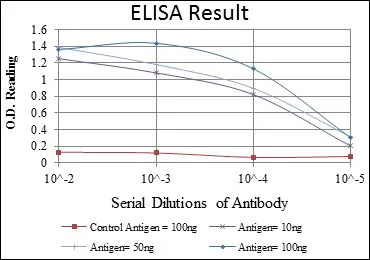
ELISA analysis of antigen using GTX60492 ERK1 antibody [1E5]. Red : Control antigen 100ng Purple : Antigen 10ng Green : Antigen 50ng Blue : Antigen 100ng
![FACS analysis of HeLa cells using GTX60492 ERK1 antibody [1E5]. Blue : ERK1 Red : negative control](https://www.genetex.com/upload/website/prouct_img/normal/GTX60492/GTX60492_20170912_FACS_w_23061123_826.webp "FACS analysis of HeLa cells using GTX60492 ERK1 antibody [1E5]. Blue : ERK1 Red : negative control")
![ICC/IF analysis of NIH3T3 cells using GTX60492 ERK1 antibody [1E5]. Green : ERK1 Blue: DRAQ5 fluorescent DNA dye Red: Actin filaments](https://www.genetex.com/upload/website/prouct_img/normal/GTX60492/GTX60492_20170912_ICCIF_w_23061123_886.webp "ICC/IF analysis of NIH3T3 cells using GTX60492 ERK1 antibody [1E5]. Green : ERK1 Blue: DRAQ5 fluorescent DNA dye Red: Actin filaments")
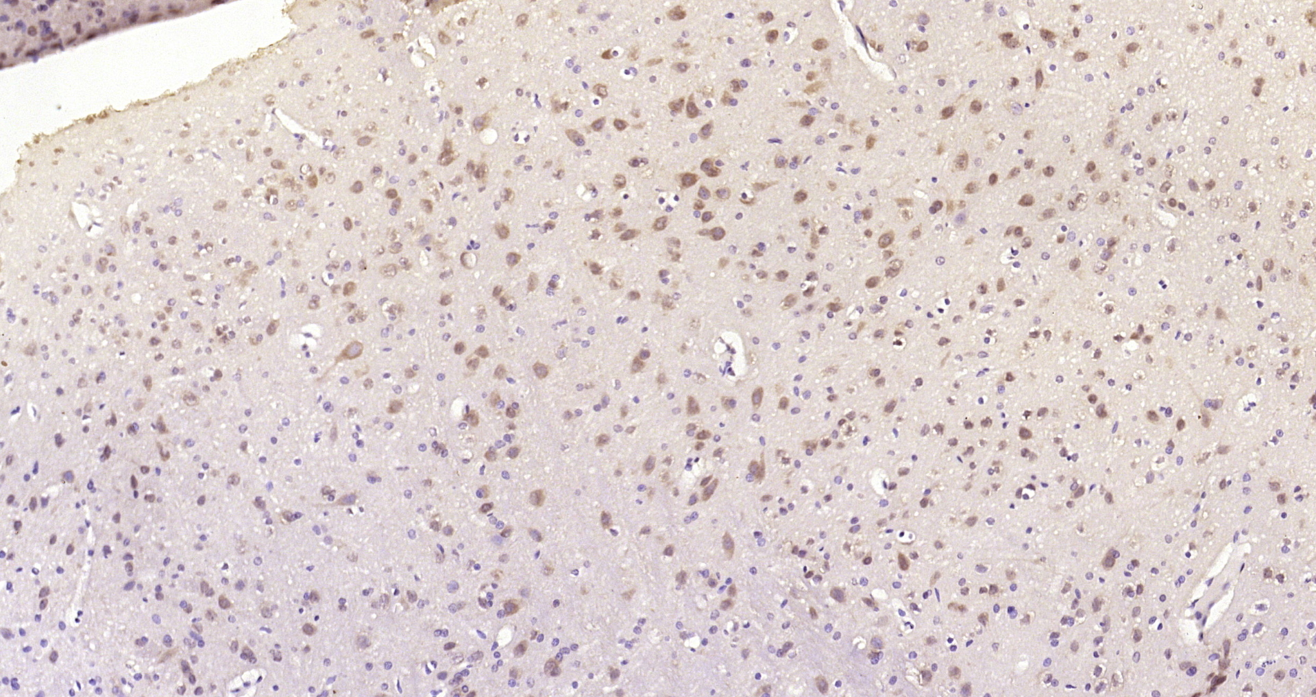
![IHC-P analysis of breast cancer tissue using GTX60492 ERK1 antibody [1E5].](https://www.genetex.com/upload/website/prouct_img/normal/GTX60492/GTX60492_20170912_IHC-P_1_w_23061123_976.webp "IHC-P analysis of breast cancer tissue using GTX60492 ERK1 antibody [1E5].")
![IHC-P analysis of bladder cancer tissue using GTX60492 ERK1 antibody [1E5].](https://www.genetex.com/upload/website/prouct_img/normal/GTX60492/GTX60492_20170912_IHC-P_w_23061123_489.webp "IHC-P analysis of bladder cancer tissue using GTX60492 ERK1 antibody [1E5].")
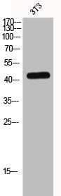
![WB analysis of human ERK1 (AA: 9-143) recombinant protein using GTX60492 ERK1 antibody [1E5].](https://www.genetex.com/upload/website/prouct_img/normal/GTX60492/GTX60492_20170912_WB_1_w_23061123_123.webp "WB analysis of human ERK1 (AA: 9-143) recombinant protein using GTX60492 ERK1 antibody [1E5].")
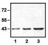
![WB analysis of Hela (1), Jurkat (2), RAW264.7 (3), HEK293 (4), K562 (5), NIH3T3 (6), Cos7 (7) and PC-12 (8) cell lysate using GTX60492 ERK1 antibody [1E5].](https://www.genetex.com/upload/website/prouct_img/normal/GTX60492/GTX60492_20170912_WB_w_23061123_228.webp "WB analysis of Hela (1), Jurkat (2), RAW264.7 (3), HEK293 (4), K562 (5), NIH3T3 (6), Cos7 (7) and PC-12 (8) cell lysate using GTX60492 ERK1 antibody [1E5].")
ELISA analysis of antigen using GTX60492 ERK1 antibody [1E5]. Red : Control antigen 100ng Purple : Antigen 10ng Green : Antigen 50ng Blue : Antigen 100ng
ERK1 antibody [1E5]
GTX60492
ApplicationsFlow Cytometry, ImmunoFluorescence, Western Blot, ELISA, ImmunoCytoChemistry, ImmunoHistoChemistry, ImmunoHistoChemistry Paraffin
Product group Antibodies
ReactivityHuman, Monkey, Mouse, Rat
TargetMAPK3
Overview
- SupplierGeneTex
- Product NameERK1 antibody [1E5]
- Delivery Days Customer9
- Application Supplier NoteWB: 1/500 - 1/2000. ICC/IF: 1/200 - 1/1000. IHC-P: 1/200 - 1/1000. FACS: 1/200 - 1/400. ELISA: 1/10000. *Optimal dilutions/concentrations should be determined by the researcher.Not tested in other applications.
- ApplicationsFlow Cytometry, ImmunoFluorescence, Western Blot, ELISA, ImmunoCytoChemistry, ImmunoHistoChemistry, ImmunoHistoChemistry Paraffin
- CertificationResearch Use Only
- ClonalityMonoclonal
- ConjugateUnconjugated
- Gene ID5595
- Target nameMAPK3
- Target descriptionmitogen-activated protein kinase 3
- Target synonymsERK-1, ERK1, ERT2, HS44KDAP, HUMKER1A, P44ERK1, P44MAPK, PRKM3, p44-ERK1, p44-MAPK, mitogen-activated protein kinase 3, MAPK 1, extracellular signal-regulated kinase 1, extracellular signal-related kinase 1, insulin-stimulated MAP2 kinase, microtubule-associated protein 2 kinase
- HostMouse
- IsotypeIgG1
- Protein IDP27361
- Protein NameMitogen-activated protein kinase 3
- Scientific DescriptionThe protein encoded by this gene is a member of the MAP kinase family. MAP kinases, also known as extracellular signal-regulated kinases (ERKs), act in a signaling cascade that regulates various cellular processes such as proliferation, differentiation, and cell cycle progression in response to a variety of extracellular signals. This kinase is activated by upstream kinases, resulting in its translocation to the nucleus where it phosphorylates nuclear targets. Alternatively spliced transcript variants encoding different protein isoforms have been described. [provided by RefSeq, Jul 2008]
- ReactivityHuman, Monkey, Mouse, Rat
- Storage Instruction-20°C or -80°C,2°C to 8°C
- UNSPSC12352203
Datasheet
Related products
Product group Antibodies
Anti-P-Erk1 (198-208aa) Antibody130-10611
ApplicationsELISA
ReactivityHuman
TargetMAPK3
- SizePrice
Product group Antibodies
MAPK3 Polyclonal AntibodyCAC13817
ApplicationsImmunoFluorescence, Western Blot, ELISA, ImmunoHistoChemistry
ReactivityMouse, Rat
TargetMAPK3
- SizePrice
Product group Antibodies
References
ApplicationsFlow Cytometry, ImmunoFluorescence, Western Blot, ELISA, ImmunoCytoChemistry, ImmunoHistoChemistry, ImmunoHistoChemistry Frozen, ImmunoHistoChemistry Paraffin
ReactivityBovine, Canine, Chicken, Equine, Guinea Pig, Human, Mouse, Porcine, Rabbit, Rat
TargetMAPK3
- SizePrice
Product group Antibodies
MAPK3 AntibodyCSB-PA002417
ApplicationsImmunoFluorescence, Western Blot, ELISA
ReactivityHuman, Mouse, Rat
TargetMAPK3
- SizePrice
![WB analysis of various samples using GTX01099 ERK1 antibody [GT1156]. Dilution : 1:1000 Loading : 25 microg](https://www.genetex.com/upload/website/prouct_img/normal/GTX01099/GTX01099_20200508_WB_w_23053121_104.webp)
Product group Antibodies
References
ERK1 antibody [GT1156]GTX01099
ApplicationsWestern Blot
ReactivityHuman, Mouse, Rat
TargetMAPK3
- SizePrice
Product group Antibodies
References
ERK1 antibodyGTX100699
ApplicationsWestern Blot
ReactivityHuman
TargetMAPK3
- SizePrice

Product group Antibodies
Anti-MAPK3 AntibodyHPA003995
ApplicationsImmunoHistoChemistry
ReactivityHuman
TargetMAPK3
- SizePrice
Product group Antibodies
ERK1 antibody [E19]GTX82557
ApplicationsImmunoPrecipitation, Western Blot
ReactivityHuman
TargetMAPK3
- SizePrice