

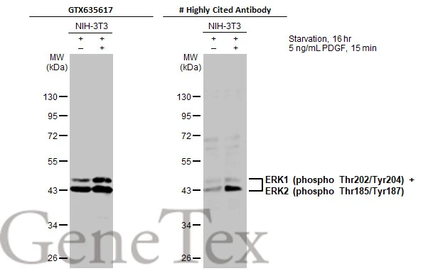
Untreated (–) and treated (+) NIH-3T3 whole cell extracts (30 μg) were separated by 10% SDS-PAGE, and the membranes were blotted with ERK1 (phospho Thr202/Tyr204) + ERK2 (phospho Thr185/Tyr187) antibody [HL173] (GTX635617) diluted at 1:500 and competitor's antibody (# Highly Cited Antibody) diluted at 1:500. The HRP-conjugated anti-rabbit IgG antibody (GTX213110-01) was used to detect the primary antibody. *The competitor is not affiliated with GeneTex and does not endorse this product.
![Untreated (–) and treated (+) A431 whole cell extracts (30 μg) were separated by 10% SDS-PAGE, and the membranes were blotted with ERK1 (phospho Thr202/Tyr204) + ERK2 (phospho Thr185/Tyr187) antibody [HL173] (GTX635617) diluted at 1:500 and competitor's antibody (# Highly Cited Antibody) diluted at 1:500. The HRP-conjugated anti-rabbit IgG antibody (GTX213110-01) was used to detect the primary antibody. *The competitor is not affiliated with GeneTex and does not endorse this product.](https://www.genetex.com/upload/website/prouct_img/normal/GTX635617/GTX635617_44711_20230217_WB_treatment_EGF_competitor_watermark_23022022_648.webp "Untreated (–) and treated (+) A431 whole cell extracts (30 μg) were separated by 10% SDS-PAGE, and the membranes were blotted with ERK1 (phospho Thr202/Tyr204) + ERK2 (phospho Thr185/Tyr187) antibody [HL173] (GTX635617) diluted at 1:500 and competitor's antibody (# Highly Cited Antibody) diluted at 1:500. The HRP-conjugated anti-rabbit IgG antibody (GTX213110-01) was used to detect the primary antibody. *The competitor is not affiliated with GeneTex and does not endorse this product.")
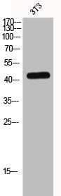
![Drosophila tissue extract (50 μg) was separated by 10% SDS-PAGE, and the membrane was blotted with ERK1 (phospho Thr202/Tyr204) + ERK2 (phospho Thr185/Tyr187) antibody [HL173] (GTX635617) diluted at 1:1000. The HRP-conjugated anti-rabbit IgG antibody (GTX213110-01) was used to detect the primary antibody, and the signal was developed with Trident ECL plus-Enhanced.](https://www.genetex.com/upload/website/prouct_img/normal/GTX635617/GTX635617_44711_20230324_WB_Drosophila_brain_23032819_703.webp "Drosophila tissue extract (50 μg) was separated by 10% SDS-PAGE, and the membrane was blotted with ERK1 (phospho Thr202/Tyr204) + ERK2 (phospho Thr185/Tyr187) antibody [HL173] (GTX635617) diluted at 1:1000. The HRP-conjugated anti-rabbit IgG antibody (GTX213110-01) was used to detect the primary antibody, and the signal was developed with Trident ECL plus-Enhanced.")
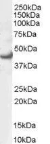
![Wild-type (WT) and ERK knockout (KO) 293T cell extracts (15 μg) were separated by 10% SDS-PAGE, and the membrane was blotted with ERK1 (phospho Thr202/Tyr204) + ERK2 (phospho Thr185/Tyr187) antibody [HL173] (GTX635617) diluted at 1:500. The HRP-conjugated anti-rabbit IgG antibody (GTX213110-01) was used to detect the primary antibody.](https://www.genetex.com/upload/website/prouct_img/normal/GTX635617/GTX635617_43978_20220211_WB_KO_watermark_w_23061202_438.webp "Wild-type (WT) and ERK knockout (KO) 293T cell extracts (15 μg) were separated by 10% SDS-PAGE, and the membrane was blotted with ERK1 (phospho Thr202/Tyr204) + ERK2 (phospho Thr185/Tyr187) antibody [HL173] (GTX635617) diluted at 1:500. The HRP-conjugated anti-rabbit IgG antibody (GTX213110-01) was used to detect the primary antibody.")
![Untreated (–) and treated (+) Rat2 whole cell extracts (30 μg) were separated by 10% SDS-PAGE, and the membrane was blotted with ERK1 (phospho Thr202/Tyr204) + ERK2 (phospho Thr185/Tyr187) antibody [HL173] (GTX635617) diluted at 1:1000. The HRP-conjugated anti-rabbit IgG antibody (GTX213110-01) was used to detect the primary antibody.](https://www.genetex.com/upload/website/prouct_img/normal/GTX635617/GTX635617_43978_20201120_WB_R_treatment_Ethacrynicacid_w_23061202_724.webp "Untreated (–) and treated (+) Rat2 whole cell extracts (30 μg) were separated by 10% SDS-PAGE, and the membrane was blotted with ERK1 (phospho Thr202/Tyr204) + ERK2 (phospho Thr185/Tyr187) antibody [HL173] (GTX635617) diluted at 1:1000. The HRP-conjugated anti-rabbit IgG antibody (GTX213110-01) was used to detect the primary antibody.")
![Untreated (–) and treated (+) Neuro2A whole cell extracts (30 μg) were separated by 10% SDS-PAGE, and the membrane was blotted with ERK1 (phospho Thr202/Tyr204) + ERK2 (phospho Thr185/Tyr187) antibody [HL173] (GTX635617) diluted at 1:1000. The HRP-conjugated anti-rabbit IgG antibody (GTX213110-01) was used to detect the primary antibody.](https://www.genetex.com/upload/website/prouct_img/normal/GTX635617/GTX635617_43978_20200619_WB_M_treatment_Ethacrynicacid_w_23061202_957.webp "Untreated (–) and treated (+) Neuro2A whole cell extracts (30 μg) were separated by 10% SDS-PAGE, and the membrane was blotted with ERK1 (phospho Thr202/Tyr204) + ERK2 (phospho Thr185/Tyr187) antibody [HL173] (GTX635617) diluted at 1:1000. The HRP-conjugated anti-rabbit IgG antibody (GTX213110-01) was used to detect the primary antibody.")
![ERK1 (phospho Thr202/Tyr204) + ERK2 (phospho Thr185/Tyr187) antibody [HL173] detects ERK1 (phospho Thr202/Tyr204) + ERK2 (phospho Thr185/Tyr187) protein at endoplasmic reticulum and cytoplasm by immunofluorescent analysis. Sample: NIH-3T3 cells were fixed in 4% paraformaldehyde at RT for 15 min. Green: ERK1 (phospho Thr202/Tyr204) + ERK2 (phospho Thr185/Tyr187) stained by ERK1 (phospho Thr202/Tyr204) + ERK2 (phospho Thr185/Tyr187) antibody [HL173] (GTX635617) diluted at 1:500. Blue: Fluoroshield with DAPI (GTX30920).](https://www.genetex.com/upload/website/prouct_img/normal/GTX635617/GTX635617_43978_20220429_ICC_IF_w_23061202_233.webp "ERK1 (phospho Thr202/Tyr204) + ERK2 (phospho Thr185/Tyr187) antibody [HL173] detects ERK1 (phospho Thr202/Tyr204) + ERK2 (phospho Thr185/Tyr187) protein at endoplasmic reticulum and cytoplasm by immunofluorescent analysis. Sample: NIH-3T3 cells were fixed in 4% paraformaldehyde at RT for 15 min. Green: ERK1 (phospho Thr202/Tyr204) + ERK2 (phospho Thr185/Tyr187) stained by ERK1 (phospho Thr202/Tyr204) + ERK2 (phospho Thr185/Tyr187) antibody [HL173] (GTX635617) diluted at 1:500. Blue: Fluoroshield with DAPI (GTX30920).")
![Untreated (–) and treated (+) A431 whole cell extracts (30 μg) were separated by 10% SDS-PAGE, and the membrane was blotted with ERK1 (phospho Thr202/Tyr204) + ERK2 (phospho Thr185/Tyr187) antibody [HL173] (GTX635617) diluted at 1:1000. The HRP-conjugated anti-rabbit IgG antibody (GTX213110-01) was used to detect the primary antibody, and the signal was developed with Trident ECL plus-Enhanced.](https://www.genetex.com/upload/website/prouct_img/normal/GTX635617/GTX635617_43978_20220128_WB_treatment_EGF_peptideblocking_w_23061202_187.webp "Untreated (–) and treated (+) A431 whole cell extracts (30 μg) were separated by 10% SDS-PAGE, and the membrane was blotted with ERK1 (phospho Thr202/Tyr204) + ERK2 (phospho Thr185/Tyr187) antibody [HL173] (GTX635617) diluted at 1:1000. The HRP-conjugated anti-rabbit IgG antibody (GTX213110-01) was used to detect the primary antibody, and the signal was developed with Trident ECL plus-Enhanced.")
![Untreated (–) and treated (+) PC-12 whole cell extracts (30 μg) were separated by 10% SDS-PAGE, and the membrane was blotted with ERK1 (phospho Thr202/Tyr204) + ERK2 (phospho Thr185/Tyr187) antibody [HL173] (GTX635617) diluted at 1:500. The HRP-conjugated anti-rabbit IgG antibody (GTX213110-01) was used to detect the primary antibody.](https://www.genetex.com/upload/website/prouct_img/normal/GTX635617/GTX635617_43978_20201009_WB_R_treatment_TPA_w_23061202_163.webp "Untreated (–) and treated (+) PC-12 whole cell extracts (30 μg) were separated by 10% SDS-PAGE, and the membrane was blotted with ERK1 (phospho Thr202/Tyr204) + ERK2 (phospho Thr185/Tyr187) antibody [HL173] (GTX635617) diluted at 1:500. The HRP-conjugated anti-rabbit IgG antibody (GTX213110-01) was used to detect the primary antibody.")
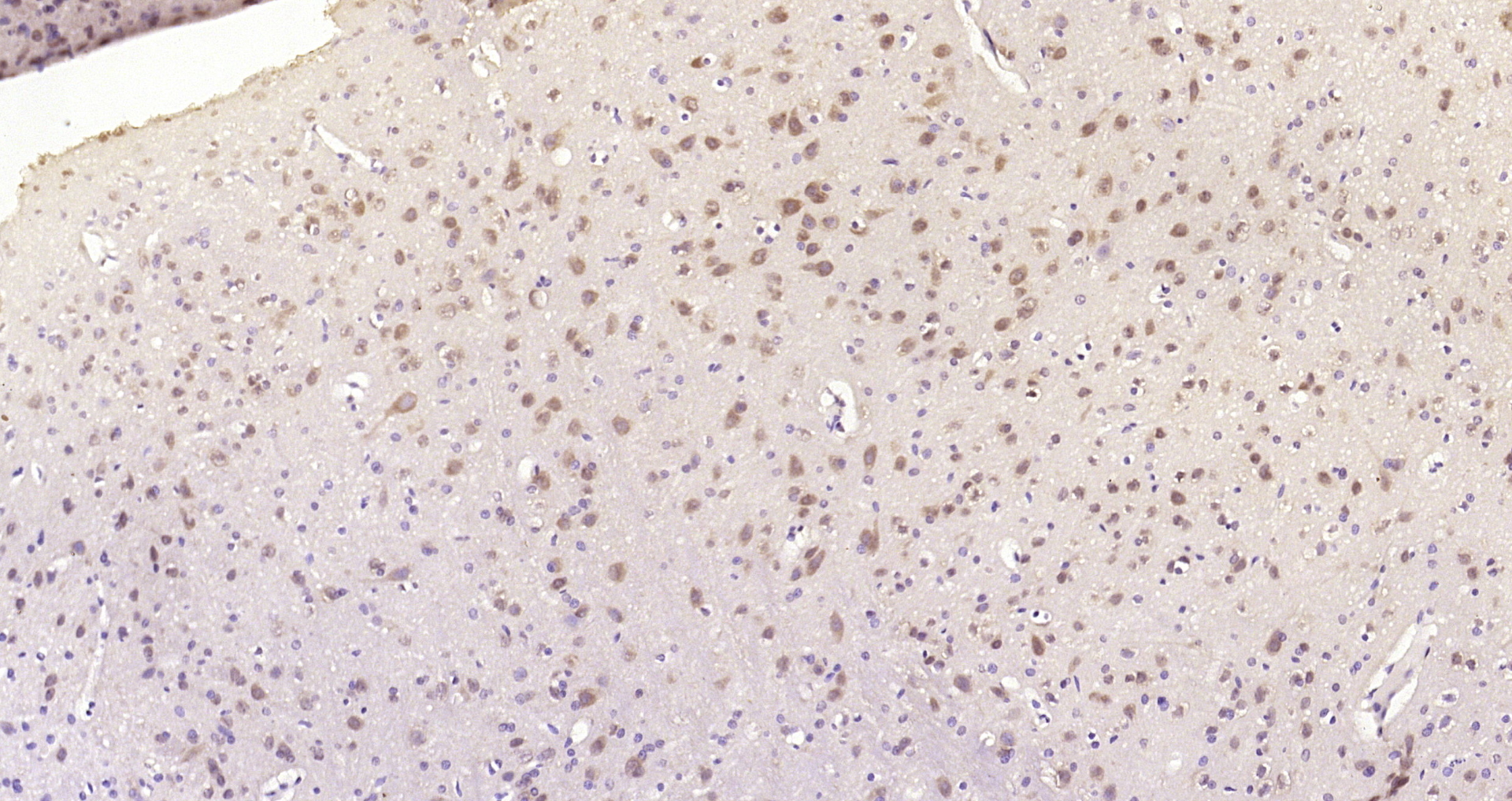
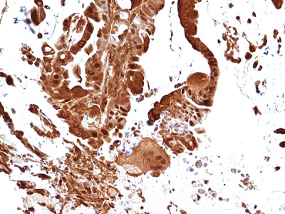
![ERK1 (phospho Thr202/Tyr204) + ERK2 (phospho Thr185/Tyr187) antibody [HL173] detects ERK1 (phospho Thr202/Tyr204) + ERK2 (phospho Thr185/Tyr187) protein at nucleus by immunohistochemical analysis. Sample: Paraffin-embedded human breast carcinoma. ERK1 (phospho Thr202/Tyr204) + ERK2 (phospho Thr185/Tyr187) stained by ERK1 (phospho Thr202/Tyr204) + ERK2 (phospho Thr185/Tyr187) antibody [HL173] (GTX635617) diluted at 1:100. Antigen Retrieval: Citrate buffer, pH 6.0, 15 min](https://www.genetex.com/upload/website/prouct_img/normal/GTX635617/GTX635617_43978_20220311_IHC-P_w_23061202_494.webp "ERK1 (phospho Thr202/Tyr204) + ERK2 (phospho Thr185/Tyr187) antibody [HL173] detects ERK1 (phospho Thr202/Tyr204) + ERK2 (phospho Thr185/Tyr187) protein at nucleus by immunohistochemical analysis. Sample: Paraffin-embedded human breast carcinoma. ERK1 (phospho Thr202/Tyr204) + ERK2 (phospho Thr185/Tyr187) stained by ERK1 (phospho Thr202/Tyr204) + ERK2 (phospho Thr185/Tyr187) antibody [HL173] (GTX635617) diluted at 1:100. Antigen Retrieval: Citrate buffer, pH 6.0, 15 min")
Untreated (–) and treated (+) NIH-3T3 whole cell extracts (30 μg) were separated by 10% SDS-PAGE, and the membranes were blotted with ERK1 (phospho Thr202/Tyr204) + ERK2 (phospho Thr185/Tyr187) antibody [HL173] (GTX635617) diluted at 1:500 and competitor's antibody (# Highly Cited Antibody) diluted at 1:500. The HRP-conjugated anti-rabbit IgG antibody (GTX213110-01) was used to detect the primary antibody. *The competitor is not affiliated with GeneTex and does not endorse this product.
ERK1 (phospho Thr202/Tyr204) + ERK2 (phospho Thr185/Tyr187) antibody [HL173]
GTX635617
ApplicationsImmunoFluorescence, Western Blot, ImmunoCytoChemistry, ImmunoHistoChemistry, ImmunoHistoChemistry Paraffin
Product group Antibodies
ReactivityDrosophila, Human, Mouse, Rat
TargetMAPK3
Overview
- SupplierGeneTex
- Product NameERK1 (phospho Thr202/Tyr204) + ERK2 (phospho Thr185/Tyr187) antibody [HL173]
- Delivery Days Customer9
- Application Supplier NoteWB: 1:500-1:3000. *Optimal dilutions/concentrations should be determined by the researcher.Not tested in other applications.
- ApplicationsImmunoFluorescence, Western Blot, ImmunoCytoChemistry, ImmunoHistoChemistry, ImmunoHistoChemistry Paraffin
- CertificationResearch Use Only
- ClonalityMonoclonal
- Clone IDHL173
- Concentration1 mg/ml
- ConjugateUnconjugated
- Gene ID5595
- Target nameMAPK3
- Target descriptionmitogen-activated protein kinase 3
- Target synonymsERK-1, ERK1, ERT2, HS44KDAP, HUMKER1A, P44ERK1, P44MAPK, PRKM3, p44-ERK1, p44-MAPK, mitogen-activated protein kinase 3, MAPK 1, extracellular signal-regulated kinase 1, extracellular signal-related kinase 1, insulin-stimulated MAP2 kinase, microtubule-associated protein 2 kinase
- HostRabbit
- IsotypeIgG
- Protein IDP27361
- Protein NameMitogen-activated protein kinase 3
- Scientific DescriptionThe protein encoded by this gene is a member of the MAP kinase family. MAP kinases, also known as extracellular signal-regulated kinases (ERKs), act in a signaling cascade that regulates various cellular processes such as proliferation, differentiation, and cell cycle progression in response to a variety of extracellular signals. This kinase is activated by upstream kinases, resulting in its translocation to the nucleus where it phosphorylates nuclear targets. Alternatively spliced transcript variants encoding different protein isoforms have been described. [provided by RefSeq, Jul 2008]
- ReactivityDrosophila, Human, Mouse, Rat
- Storage Instruction-20°C or -80°C,2°C to 8°C
- UNSPSC12352203
References
- Watanabe M, Kawaguchi K, Nakamura Y, et al. GIF-2209, an Oxindole Derivative, Accelerates Melanogenesis and Melanosome Secretion via the Modification of Lysosomes in B16F10 Mouse Melanoma Cells. Molecules. 2021,27(1). doi: 10.3390/molecules27010177Read this paper
- Chung WP, Huang WL, Liao WA, et al. FTY720 in resistant human epidermal growth factor receptor 2-positive breast cancer. Sci Rep. 2022,12(1):241. doi: 10.1038/s41598-021-04328-yRead this paper
- Tsai HJ, Shih CC, Chang KY, et al. Angiotensin-(1-7) treatment blocks lipopolysaccharide-induced organ damage, platelet dysfunction, and IL-6 and nitric oxide production in rats. Sci Rep. 2021,11(1):610. doi: 10.1038/s41598-020-79902-xRead this paper
- Liu JF, Chi MC, Lin CY, et al. PM2.5 facilitates IL-6 production in human osteoarthritis synovial fibroblasts via ASK1 activation. J Cell Physiol. 2021,236(3):2205-2213. doi: 10.1002/jcp.30009Read this paper
Datasheet
Related products
Product group Antibodies
Anti-P-Erk1 (198-208aa) Antibody130-10611
ApplicationsELISA
ReactivityHuman
TargetMAPK3
- SizePrice
Product group Antibodies
MAPK3 Polyclonal AntibodyCAC13817
ApplicationsImmunoFluorescence, Western Blot, ELISA, ImmunoHistoChemistry
ReactivityMouse, Rat
TargetMAPK3
- SizePrice
Product group Antibodies
References
ApplicationsFlow Cytometry, ImmunoFluorescence, Western Blot, ELISA, ImmunoCytoChemistry, ImmunoHistoChemistry, ImmunoHistoChemistry Frozen, ImmunoHistoChemistry Paraffin
ReactivityBovine, Canine, Chicken, Equine, Guinea Pig, Human, Mouse, Porcine, Rabbit, Rat
TargetMAPK3
- SizePrice
Product group Antibodies
MAPK3 AntibodyCSB-PA002417
ApplicationsImmunoFluorescence, Western Blot, ELISA
ReactivityHuman, Mouse, Rat
TargetMAPK3
- SizePrice
Product group Antibodies

MAPK3 / ERK1 Antibody (clone 1C11)LS-C766423
ApplicationsImmunoHistoChemistry, ImmunoHistoChemistry Paraffin
ReactivityHuman, Mouse, Rat
TargetMAPK3
- SizePrice
Product group Antibodies
ERK1 antibody, N-termGTX89563
ApplicationsWestern Blot
ReactivityHuman
TargetMAPK3
- SizePrice
Product group Antibodies
Anti-MAPK3 AntibodyA95846
ApplicationsWestern Blot, ELISA, ImmunoHistoChemistry
ReactivityHuman, Mouse, Rat
- SizePrice
Product group Antibodies
anti-p44 MAPK (Erk1), Rabbit Monoclonal (RM470)REV-31-1362-00
ApplicationsWestern Blot, ImmunoHistoChemistry
ReactivityHuman
TargetMAPK3
- SizePrice