

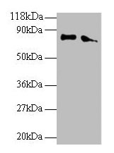
Western blot All lanes: ERP29 antibody at 2microg/ml Lane 1: EC109 whole cell lysate Lane 2: 293T whole cell lysate Secondary Goat polyclonal to rabbit IgG at 1/15000 dilution Predicted band size: 29, 6 kDa Observed band size: 80 kDa

Western blot All lanes: ERP29 antibody at 2microg/ml Lane 1: EC109 whole cell lysate Lane 2: 293T whole cell lysate Secondary Goat polyclonal to rabbit IgG at 1/15000 dilution Predicted band size: 29, 6 kDa Observed band size: 80 kDa
ERP29 Antibody
CSB-PA05344A0RB
ApplicationsWestern Blot, ELISA, ImmunoHistoChemistry
Product group Antibodies
ReactivityHuman
TargetERP29
Overview
- SupplierCusabio
- Product NameERP29 Antibody
- Delivery Days Customer20
- ApplicationsWestern Blot, ELISA, ImmunoHistoChemistry
- CertificationResearch Use Only
- ClonalityPolyclonal
- ConjugateUnconjugated
- Gene ID10961
- Target nameERP29
- Target descriptionendoplasmic reticulum protein 29
- Target synonymsC12orf8, ERp28, ERp31, HEL-S-107, PDI-DB, PDIA9, endoplasmic reticulum resident protein 29, endoplasmic reticulum lumenal protein ERp28, endoplasmic reticulum resident protein 28, endoplasmic reticulum resident protein 31, epididymis secretory protein Li 107, protein disulfide isomerase family A, member 9
- HostRabbit
- IsotypeIgG
- Protein IDP30040
- Protein NameEndoplasmic reticulum resident protein 29
- Scientific DescriptionProper protein folding and post-translational modifications are essential for secretory protein export out of the endoplasmic reticulum. This task is accomplished by chaperone proteins such as protein disulfide isomerase (PDI), GRP94, and BiP. A recently characterized protein, designated ERp29, is closely related to these chaperone proteins and appears to be upregulated during ER stress conditions. ERp29 is a soluble 259-residue protein that is localized to the lumen of the endoplasmic reticulum in all mammalian cells. Research has shown that there are two primary domains within ERp29. The first is the C-terminal region that is a novel, all helical, fold that is most likely involved with ERp29 retention to the ER. The second is the N-terminal region that resembles that of PDIs thioredoxin module. The protein shows sequence similarity to the protein disulfide isomerase family. However, it lacks the thioredoxin motif characteristic of this family, suggesting that this protein does not function as a disulfide isomerase. The protein dimerizes and is thought to play a role in the processing of secretory proteins within the ER.
- ReactivityHuman
- Storage Instruction-20°C or -80°C
- UNSPSC41116161
Related products
Product group Antibodies
Anti-ERP29 AntibodyA38053
ApplicationsWestern Blot, ImmunoHistoChemistry
ReactivityHuman, Mouse, Rat
- SizePrice

Product group Antibodies
Anti-ERP29 Antibody Picoband(r)A03621-2-CARRIER-FREE
ApplicationsFlow Cytometry, ImmunoFluorescence, Western Blot, ELISA, ImmunoCytoChemistry, ImmunoHistoChemistry
ReactivityHuman, Monkey, Mouse, Rat
TargetERP29
- SizePrice
Product group Antibodies
Anti-ERP29 Antibody144-07959
ApplicationsWestern Blot
ReactivityHuman, Mouse, Rat
TargetERP29
- SizePrice
Product group Antibodies
ERp29 Recombinant Antibody, AbBy Fluor-555 ConjugatedBSM-61892R-BF555
ApplicationsImmunoFluorescence, Western Blot
ReactivityHuman, Mouse, Rat
TargetERP29
- SizePrice
Product group Antibodies
References

Goat anti-ERP29EB08977
ApplicationsImmunoFluorescence, Western Blot, ELISA
ReactivityBovine, Canine, Human, Porcine, Rat
TargetERP29
- SizePrice
Product group Antibodies
ERP29 Polyclonal AntibodyCAC14074
ApplicationsWestern Blot, ELISA, ImmunoHistoChemistry
TargetERP29
- SizePrice
Product group Antibodies

ERP29 AntibodyLS-C403137
ApplicationsWestern Blot, ELISA, ImmunoHistoChemistry
ReactivityHuman, Mouse, Rat
TargetERP29
- SizePrice
Product group Antibodies
ERP29 antibodyGTX102225
ApplicationsImmunoFluorescence, Western Blot, ImmunoCytoChemistry, ImmunoHistoChemistry, ImmunoHistoChemistry Paraffin
ReactivityHuman, Mouse, Rat
TargetERP29
- SizePrice

Product group Antibodies
Anti-ERP29 AntibodyHPA039363
ApplicationsWestern Blot, ImmunoHistoChemistry
ReactivityHuman
TargetERP29
- SizePrice