

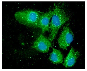
ICC/IF analysis of Hep3B cells using GTX57663 ERp57 antibody. Blue: DAPI Green: Primary antibody Dilution: 1:100

ICC/IF analysis of Hep3B cells using GTX57663 ERp57 antibody. Blue: DAPI Green: Primary antibody Dilution: 1:100
ERp57 antibody [AT9E9]
GTX57663
ApplicationsImmunoFluorescence, Western Blot, ImmunoCytoChemistry
Product group Antibodies
ReactivityHuman, Mouse
TargetPDIA3
Overview
- SupplierGeneTex
- Product NameERp57 antibody [AT9E9]
- Delivery Days Customer9
- ApplicationsImmunoFluorescence, Western Blot, ImmunoCytoChemistry
- CertificationResearch Use Only
- ClonalityMonoclonal
- Clone IDAT9E9
- Concentration1 mg/ml
- ConjugateUnconjugated
- Gene ID2923
- Target namePDIA3
- Target descriptionprotein disulfide isomerase family A member 3
- Target synonymsER60, ERp57, ERp60, ERp61, GRP57, GRP58, HEL-S-269, HEL-S-93n, HsT17083, P58, PI-PLC, protein disulfide-isomerase A3, 58 kDa glucose-regulated protein, 58 kDa microsomal protein, ER protein 57, ER protein 60, disulfide isomerase ER-60, endoplasmic reticulum P58, endoplasmic reticulum resident protein 57, endoplasmic reticulum resident protein 60, epididymis secretory protein Li 269, epididymis secretory sperm binding protein Li 93n, glucose regulated protein, 58kDa, phospholipase C-alpha, protein disulfide isomerase-associated 3
- HostMouse
- IsotypeIgG2a
- Protein IDP30101
- Protein NameProtein disulfide-isomerase A3
- Scientific DescriptionThis gene encodes a protein of the endoplasmic reticulum that interacts with lectin chaperones calreticulin and calnexin to modulate folding of newly synthesized glycoproteins. The protein was once thought to be a phospholipase; however, it has been demonstrated that the protein actually has protein disulfide isomerase activity. It is thought that complexes of lectins and this protein mediate protein folding by promoting formation of disulfide bonds in their glycoprotein substrates. This protein also functions as a molecular chaperone that prevents the formation of protein aggregates. [provided by RefSeq, Dec 2016]
- ReactivityHuman, Mouse
- Storage Instruction-20°C or -80°C,2°C to 8°C
- UNSPSC12352203
Datasheet
Related products
Product group Antibodies
Anti-PDIA3 Antibody144-01085
ApplicationsImmunoFluorescence, Western Blot, ImmunoHistoChemistry
ReactivityHuman, Mouse, Rat
TargetPDIA3
- SizePrice

Product group Antibodies
Anti-PDIA3 AntibodyAMAB90988
ApplicationsWestern Blot, ImmunoCytoChemistry, ImmunoHistoChemistry
ReactivityHuman
TargetPDIA3
- SizePrice
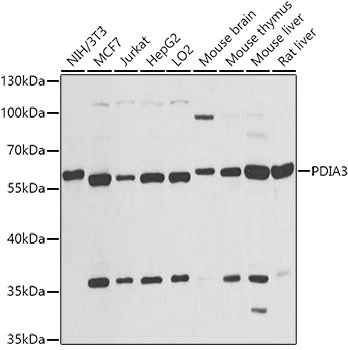
![Various whole cell extracts (30 μg) were separated by 10% SDS-PAGE, and the membrane was blotted with ERp57 antibody [C3], C-term (GTX100297) diluted at 1:10000. The HRP-conjugated anti-rabbit IgG antibody (GTX213110-01) was used to detect the primary antibody.](https://www.genetex.com/upload/website/prouct_img/normal/GTX100297/GTX100297_39422_20190624_WB_R_w_23060100_776.webp)
Product group Antibodies
References
ERp57 antibody [C3], C-termGTX100297
ApplicationsImmunoFluorescence, Western Blot, ImmunoCytoChemistry, ImmunoHistoChemistry, ImmunoHistoChemistry Paraffin
ReactivityHuman, Mouse, Rat
TargetPDIA3
- SizePrice
![ERp57 antibody detects ERp57 protein at endoplasmic reticulum by immunofluorescent analysis. Sample: HeLa cells were fixed in 4% paraformaldehyde at RT for 15 min. Green: ERp57 protein stained by ERp57 antibody (GTX113719) diluted at 1:1000. Red: alpha Tubulin, a cytoskeleton marker, stained by alpha Tubulin antibody [GT114] (GTX628802) diluted at 1:1000. Blue: Hoechst 33342 staining.](https://www.genetex.com/upload/website/prouct_img/normal/GTX113719/GTX113719_40471_20150410_IFA_w_23060501_406.webp)
Product group Antibodies
References
ERp57 antibodyGTX113719
ApplicationsImmunoFluorescence, Western Blot, ImmunoCytoChemistry, ImmunoHistoChemistry, ImmunoHistoChemistry Paraffin
ReactivityHuman, Mouse, Rat
TargetPDIA3
- SizePrice
Product group Antibodies
ERp57 Recombinant Antibody, AbBy Fluor-350 ConjugatedBSM-61463R-BF350
ApplicationsImmunoFluorescence, Western Blot
ReactivityHuman
TargetPDIA3
- SizePrice
Product group Antibodies
PDIA3 AntibodyCSB-PA005150
ApplicationsWestern Blot, ELISA
ReactivityHuman, Mouse, Rat
TargetPDIA3
- SizePrice

Product group Antibodies
Anti-PDIA3 AntibodyA97355
ApplicationsWestern Blot, ELISA
ReactivityHuman, Mouse, Rat
- SizePrice
Product group Antibodies
ERp57 antibodyGTX17012
ApplicationsWestern Blot
ReactivityHuman, Mouse
TargetPDIA3
- SizePrice
Product group Antibodies
ERp57 antibodyGTX32592
ApplicationsWestern Blot, ImmunoHistoChemistry, ImmunoHistoChemistry Paraffin
ReactivityHuman, Mouse, Rat
TargetPDIA3
- SizePrice