

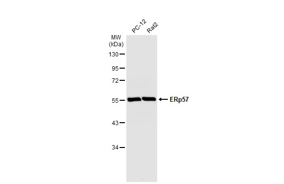
Various whole cell extracts (30 μg) were separated by 10% SDS-PAGE, and the membrane was blotted with ERp57 antibody [C3], C-term (GTX100297) diluted at 1:10000. The HRP-conjugated anti-rabbit IgG antibody (GTX213110-01) was used to detect the primary antibody.
 A: NIH-3T3 B: JC C: BCL-1 7.5% SDS PAGE GTX100297 diluted at 1:1000")
![ERp57 antibody [C3], C-term detects ERp57 protein at cytosol on mouse intestine by immunohistochemical analysis. Sample: Paraffin-embedded mouse intestine. ERp57 antibody [C3], C-term (GTX100297) dilution: 1:500.
Antigen Retrieval: Trilogy? (EDTA based, pH 8.0) buffer, 15min](https://www.genetex.com/upload/website/prouct_img/normal/GTX100297/GTX100297_39422_IHC_M_w_23060100_692.webp "ERp57 antibody [C3], C-term detects ERp57 protein at cytosol on mouse intestine by immunohistochemical analysis. Sample: Paraffin-embedded mouse intestine. ERp57 antibody [C3], C-term (GTX100297) dilution: 1:500.
Antigen Retrieval: Trilogy? (EDTA based, pH 8.0) buffer, 15min")
![ERp57 antibody [C3], C-term detects ERp57 protein at cytosol on mouse intestine by immunohistochemical analysis. Sample: Paraffin-embedded mouse intestine. ERp57 antibody [C3], C-term (GTX100297) dilution: 1:500.
Antigen Retrieval: Trilogy? (EDTA based, pH 8.0) buffer, 15min](https://www.genetex.com/upload/website/prouct_img/normal/GTX100297/GTX100297_39422_IHC_M_2_w_23060100_982.webp "ERp57 antibody [C3], C-term detects ERp57 protein at cytosol on mouse intestine by immunohistochemical analysis. Sample: Paraffin-embedded mouse intestine. ERp57 antibody [C3], C-term (GTX100297) dilution: 1:500.
Antigen Retrieval: Trilogy? (EDTA based, pH 8.0) buffer, 15min")
![ERp57 antibody [C3], C-term detects ERp57 protein at cytoplasm by immunofluorescent analysis. Sample: HeLa cells were fixed in 4% paraformaldehyde at RT for 15 min. Green: ERp57 protein stained by ERp57 antibody [C3], C-term (GTX100297) diluted at 1:1000. Red: alpha Tubulin, a cytoskeleton marker, stained by alpha Tubulin antibody [GT114] (GTX628802) diluted at 1:1000. Blue: Hoechst 33342 staining.](https://www.genetex.com/upload/website/prouct_img/normal/GTX100297/GTX100297_39422_20150410_IFA_w_23060100_451.webp "ERp57 antibody [C3], C-term detects ERp57 protein at cytoplasm by immunofluorescent analysis. Sample: HeLa cells were fixed in 4% paraformaldehyde at RT for 15 min. Green: ERp57 protein stained by ERp57 antibody [C3], C-term (GTX100297) diluted at 1:1000. Red: alpha Tubulin, a cytoskeleton marker, stained by alpha Tubulin antibody [GT114] (GTX628802) diluted at 1:1000. Blue: Hoechst 33342 staining.")
 antibody at 1:500 dilution.
Antigen Retrieval: Trilogy? (EDTA based, pH 8.0) buffer, 15min")
![ERp57 antibody [C3], C-term detects PDIA3 protein at cytoplasm by immunofluorescent analysis. Sample: MCF-7 cells were fixed in ice-cold MeOH for 5 min. Green: PDIA3 protein stained by ERp57 antibody [C3], C-term (GTX100297) diluted at 1:500. Blue: Hoechst 33342 staining.](https://www.genetex.com/upload/website/prouct_img/normal/GTX100297/GTX100297_39428_IFA_w_23060100_439.webp "ERp57 antibody [C3], C-term detects PDIA3 protein at cytoplasm by immunofluorescent analysis. Sample: MCF-7 cells were fixed in ice-cold MeOH for 5 min. Green: PDIA3 protein stained by ERp57 antibody [C3], C-term (GTX100297) diluted at 1:500. Blue: Hoechst 33342 staining.")
![ERp57 antibody [C3], C-term detects ERp57 protein at cytosol on rat middle brain by immunohistochemical analysis. Sample: Paraffin-embedded rat middle brain. ERp57 antibody [C3], C-term (GTX100297) dilution: 1:500.
Antigen Retrieval: Trilogy? (EDTA based, pH 8.0) buffer, 15min](https://www.genetex.com/upload/website/prouct_img/normal/GTX100297/GTX100297_39422_IHC_R_w_23060100_939.webp "ERp57 antibody [C3], C-term detects ERp57 protein at cytosol on rat middle brain by immunohistochemical analysis. Sample: Paraffin-embedded rat middle brain. ERp57 antibody [C3], C-term (GTX100297) dilution: 1:500.
Antigen Retrieval: Trilogy? (EDTA based, pH 8.0) buffer, 15min")
![Various whole cell extracts (30 μg) were separated by 10% SDS-PAGE, and the membranes were blotted with ERp57 antibody [C3], C-term (GTX100297) diluted at 1:5000 and competitor's antibody diluted at 1:5000. The HRP-conjugated anti-rabbit IgG antibody (GTX213110-01) was used to detect the primary antibody. *The competitor is not affiliated with GeneTex and does not endorse this product.](https://www.genetex.com/upload/website/prouct_img/normal/GTX100297/GTX100297_39422_20200117_WB_competitor_watermark_w_23060100_150.webp "Various whole cell extracts (30 μg) were separated by 10% SDS-PAGE, and the membranes were blotted with ERp57 antibody [C3], C-term (GTX100297) diluted at 1:5000 and competitor's antibody diluted at 1:5000. The HRP-conjugated anti-rabbit IgG antibody (GTX213110-01) was used to detect the primary antibody. *The competitor is not affiliated with GeneTex and does not endorse this product.")
 A: H1299 B: Hela C: Hep G2 (GTX27900) 7.5% SDS PAGE GTX100297 diluted at 1:10000")
Various whole cell extracts (30 μg) were separated by 10% SDS-PAGE, and the membrane was blotted with ERp57 antibody [C3], C-term (GTX100297) diluted at 1:10000. The HRP-conjugated anti-rabbit IgG antibody (GTX213110-01) was used to detect the primary antibody.
ERp57 antibody [C3], C-term
GTX100297
ApplicationsImmunoFluorescence, Western Blot, ImmunoCytoChemistry, ImmunoHistoChemistry, ImmunoHistoChemistry Paraffin
Product group Antibodies
ReactivityHuman, Mouse, Rat
TargetPDIA3
Overview
- SupplierGeneTex
- Product NameERp57 antibody [C3], C-term
- Delivery Days Customer9
- Application Supplier NoteWB: 1:1000-1:10000. ICC/IF: 1:100-1:1000. IHC-P: 1:100-1:1000. *Optimal dilutions/concentrations should be determined by the researcher.Not tested in other applications.
- ApplicationsImmunoFluorescence, Western Blot, ImmunoCytoChemistry, ImmunoHistoChemistry, ImmunoHistoChemistry Paraffin
- CertificationResearch Use Only
- ClonalityPolyclonal
- Concentration1 mg/ml
- ConjugateUnconjugated
- Gene ID2923
- Target namePDIA3
- Target descriptionprotein disulfide isomerase family A member 3
- Target synonymsER60, ERp57, ERp60, ERp61, GRP57, GRP58, HEL-S-269, HEL-S-93n, HsT17083, P58, PI-PLC, protein disulfide-isomerase A3, 58 kDa glucose-regulated protein, 58 kDa microsomal protein, ER protein 57, ER protein 60, disulfide isomerase ER-60, endoplasmic reticulum P58, endoplasmic reticulum resident protein 57, endoplasmic reticulum resident protein 60, epididymis secretory protein Li 269, epididymis secretory sperm binding protein Li 93n, glucose regulated protein, 58kDa, phospholipase C-alpha, protein disulfide isomerase-associated 3
- HostRabbit
- IsotypeIgG
- Protein IDP30101
- Protein NameProtein disulfide-isomerase A3
- Scientific DescriptionThis gene encodes a protein of the endoplasmic reticulum that interacts with lectin chaperones calreticulin and calnexin to modulate folding of newly synthesized glycoproteins. The protein was once thought to be a phospholipase; however, it has been demonstrated that the protein actually has protein disulfide isomerase activity. It is thought that complexes of lectins and this protein mediate protein folding by promoting formation of disulfide bonds in their glycoprotein substrates. [provided by RefSeq]
- ReactivityHuman, Mouse, Rat
- Storage Instruction-20°C or -80°C,2°C to 8°C
- UNSPSC41116161
Datasheet
Related products

Product group Antibodies
Anti-PDIA3 AntibodyA97355
ApplicationsWestern Blot, ELISA
ReactivityHuman, Mouse, Rat
- SizePrice
Product group Antibodies
Anti-PDIA3 Antibody144-01085
ApplicationsImmunoFluorescence, Western Blot, ImmunoHistoChemistry
ReactivityHuman, Mouse, Rat
TargetPDIA3
- SizePrice

Product group Antibodies
Anti-PDIA3 AntibodyAMAB90988
ApplicationsWestern Blot, ImmunoCytoChemistry, ImmunoHistoChemistry
ReactivityHuman
TargetPDIA3
- SizePrice
Product group Antibodies
ERp57 Recombinant Antibody, AbBy Fluor-350 ConjugatedBSM-61463R-BF350
ApplicationsImmunoFluorescence, Western Blot
ReactivityHuman
TargetPDIA3
- SizePrice
Product group Antibodies

ApplicationsWestern Blot, ELISA
ReactivityBovine, Human, Mouse, Rat
TargetPDIA3
- SizePrice
Product group Antibodies
ApplicationsImmunoPrecipitation, Western Blot, ImmunoCytoChemistry, ImmunoHistoChemistry
ReactivityMouse, Porcine, Rat
TargetPDIA3
- SizePrice
Product group Antibodies
PDIA3 AntibodyCSB-PA005150
ApplicationsWestern Blot, ELISA
ReactivityHuman, Mouse, Rat
TargetPDIA3
- SizePrice
Product group Antibodies
ERp57 antibody [MaP.Erp57]GTX13506
ApplicationsImmunoPrecipitation, Western Blot
ReactivityCanine, Hamster, Human, Monkey, Mouse, Porcine
TargetPDIA3
- SizePrice
Product group Antibodies
ERp57 antibodyGTX17012
ApplicationsWestern Blot
ReactivityHuman, Mouse
TargetPDIA3
- SizePrice
Product group Antibodies

PDIA3 / ERp57 AntibodyLS-C331251
ApplicationsWestern Blot, ImmunoHistoChemistry
ReactivityHuman, Mouse, Rat
TargetPDIA3
- SizePrice