

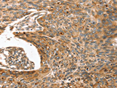
The image on the left is immunohistochemistry of paraffin-embedded Human esophagus cancer tissue using CSB-PA215261(ERVW-1 Antibody) at dilution 1/45, on the right is treated with synthetic peptide. (Original magnification: x200)
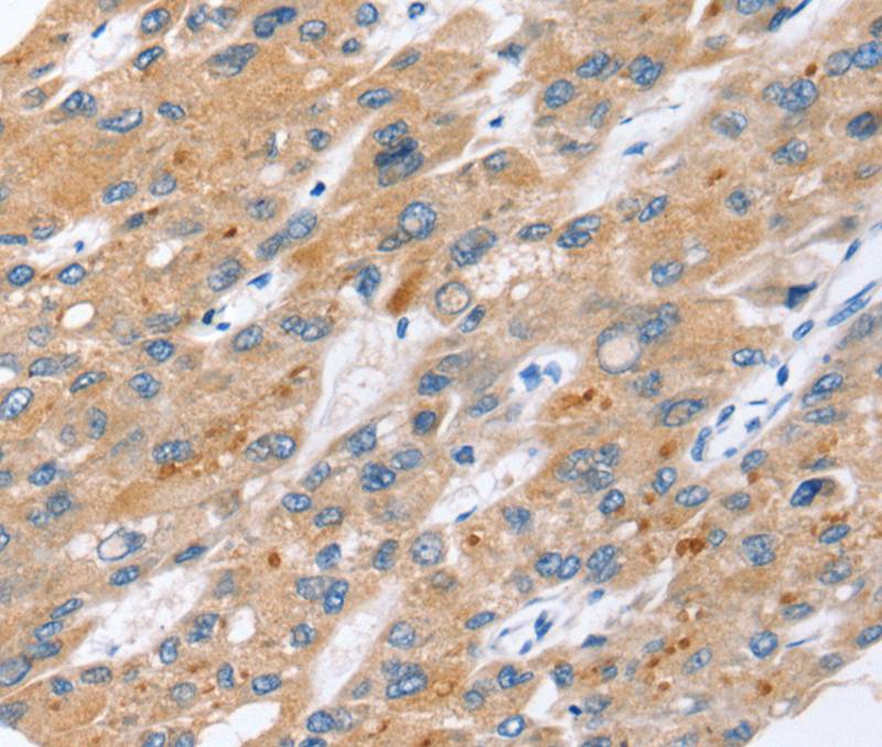
The image on the left is immunohistochemistry of paraffin-embedded Human esophagus cancer tissue using CSB-PA215261(ERVW-1 Antibody) at dilution 1/45, on the right is treated with synthetic peptide. (Original magnification: x200)
ERVW-1 Antibody
CSB-PA215261
ApplicationsELISA, ImmunoHistoChemistry
Product group Antibodies
ReactivityHuman
TargetERVW-1
Overview
- SupplierCusabio
- Product NameERVW-1 Antibody
- Delivery Days Customer20
- ApplicationsELISA, ImmunoHistoChemistry
- CertificationResearch Use Only
- ClonalityPolyclonal
- ConjugateUnconjugated
- Gene ID30816
- Target nameERVW-1
- Target descriptionendogenous retrovirus group W member 1, envelope
- Target synonymsENV, ENVW, ERVWE1, HERV-7q, HERV-W-ENV, HERV7Q, HERVW, HERVWENV, syncytin-1, HERV-7q envelope protein, HERV-W Env glycoprotein, HERV-W envelope protein, HERV-W_7q21.2 provirus ancestral Env polyprotein, HERV-tryptophan envelope protein, endogenous retroviral family W, env(C7), member 1, endogenous retrovirus group W, member 1, envelope glycoprotein, envelope polyprotein gPr73, enverin, human endogenous retrovirus W envC7-1 envelope protein
- HostRabbit
- IsotypeIgG
- Protein IDQ9UQF0
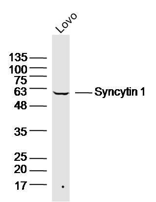
- Protein NameSyncytin-1
- Scientific DescriptionMany different human endogenous retrovirus (HERV) families are expressed in normal placental tissue at high levels, suggesting that HERVs are functionally important in reproduction. This gene is part of an HERV provirus on chromosome 7 that has inactivating mutations in the gag and pol genes. This gene is the envelope glycoprotein gene which appears to have been selectively preserved. The genes protein product is expressed in the placental syncytiotrophoblast and is involved in fusion of the cytotrophoblast cells to form the syncytial layer of the placenta.
- ReactivityHuman
- Storage Instruction-20°C or -80°C
- UNSPSC41116161
Related products
Product group Antibodies
Anti-ERVW-1 AntibodyA45628
ApplicationsImmunoHistoChemistry
ReactivityHuman
- SizePrice
Product group Antibodies
Anti-ERVW-1 Antibody144-64458
ApplicationsWestern Blot
ReactivityHuman, Mouse
TargetERVW-1
- SizePrice
Product group Antibodies
Ervw-1 Polyclonal AntibodyCAC09941
ApplicationsImmunoFluorescence, ELISA, ImmunoHistoChemistry
TargetERVW-1
- SizePrice
Product group Antibodies
References
Syncytin 1 Polyclonal AntibodyBS-2962R
ApplicationsWestern Blot, ELISA
ReactivityHuman, Mouse
TargetERVW-1
- SizePrice
Product group Antibodies

ApplicationsELISA, ImmunoHistoChemistry
ReactivityHuman
TargetERVW-1
- SizePrice
Product group Antibodies
References
ERVWE1 antibody, N-termGTX46373
ApplicationsWestern Blot
ReactivityHamster, Human
TargetERVW-1
- SizePrice

Product group Antibodies
Anti-ERVW-1 Antibody Picoband(r)PB9622-CARRIER-FREE
ApplicationsWestern Blot
ReactivityHuman
TargetERVW-1
- SizePrice