

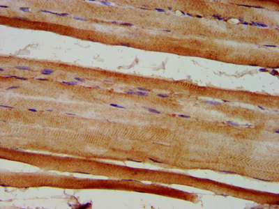
IHC image of CSB-PA007909LA01HU diluted at 1:600 and staining in paraffin-embedded human skeletal muscle tissue performed on a Leica BondTM system. After dewaxing and hydration, antigen retrieval was mediated by high pressure in a citrate buffer (pH 6.0). Section was blocked with 10% normal goat serum 30min at RT. Then primary antibody (1% BSA) was incubated at 4°C overnight. The primary is detected by a biotinylated secondary antibody and visualized using an HRP conjugated SP system.
.")
IHC image of CSB-PA007909LA01HU diluted at 1:600 and staining in paraffin-embedded human skeletal muscle tissue performed on a Leica BondTM system. After dewaxing and hydration, antigen retrieval was mediated by high pressure in a citrate buffer (pH 6.0). Section was blocked with 10% normal goat serum 30min at RT. Then primary antibody (1% BSA) was incubated at 4°C overnight. The primary is detected by a biotinylated secondary antibody and visualized using an HRP conjugated SP system.
EYA4 Antibody
CSB-PA007909LA01HU
ApplicationsImmunoFluorescence, ELISA, ImmunoHistoChemistry
Product group Antibodies
ReactivityHuman
TargetEYA4
Overview
- SupplierCusabio
- Product NameEYA4 Antibody
- Delivery Days Customer20
- ApplicationsImmunoFluorescence, ELISA, ImmunoHistoChemistry
- CertificationResearch Use Only
- ClonalityPolyclonal
- ConjugateUnconjugated
- Gene ID2070
- Target nameEYA4
- Target descriptionEYA transcriptional coactivator and phosphatase 4
- Target synonymsCMD1J, DFNA10, protein phosphatase EYA4, dJ78N10.1 (eyes absent), eyes absent homolog 4, eyes absent-like protein 4
- HostRabbit
- IsotypeIgG
- Protein IDO95677
- Protein NameProtein phosphatase EYA4
- Scientific DescriptionTyrosine phosphatase that specifically dephosphorylates Tyr-142 of histone H2AX (H2AXY142ph). Tyr-142 phosphorylation of histone H2AX plays a central role in DNA repair and acts as a mark that distinguishes between apoptotic and repair responses to genotoxic stress. Promotes efficient DNA repair by dephosphorylating H2AX, promoting the recruitment of DNA repair complexes containing MDC1. Its function as histone phosphatase probably explains its role in transcription regulation during organogenesis. May be involved in development of the eye (By similarity).
- ReactivityHuman
- Storage Instruction-20°C or -80°C
- UNSPSC41116161
Related products
Product group Antibodies
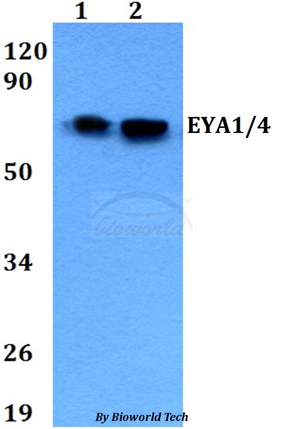
Anti-EYA1/4 AntibodyA28866
ApplicationsWestern Blot
ReactivityHuman, Rat
- SizePrice

Product group Antibodies
Anti-EYA4 Antibody Picoband(r)A04516-3-CARRIER-FREE
ApplicationsFlow Cytometry, Western Blot, ELISA, ImmunoHistoChemistry
ReactivityHuman, Monkey, Rat
TargetEYA4
- SizePrice
Product group Antibodies
EYA4 Polyclonal AntibodyBS-13126R
ApplicationsImmunoFluorescence, ELISA, ImmunoCytoChemistry, ImmunoHistoChemistry, ImmunoHistoChemistry Frozen, ImmunoHistoChemistry Paraffin
ReactivityBovine, Canine, Equine, Human, Mouse, Porcine, Rat, Sheep
TargetEYA4
- SizePrice
Product group Antibodies

Goat anti-EYA4EB06993
ApplicationsELISA, ImmunoHistoChemistry
ReactivityHuman, Mouse
TargetEYA4
- SizePrice
Product group Antibodies

EYA4 AntibodyLS-C411172
ApplicationsFlow Cytometry, Western Blot, ImmunoHistoChemistry
ReactivityHuman
TargetEYA4
- SizePrice

Product group Antibodies
Anti-EYA4 AntibodyHPA004805
ApplicationsWestern Blot, ImmunoCytoChemistry, ImmunoHistoChemistry
ReactivityHuman
TargetEYA4
- SizePrice
Product group Antibodies
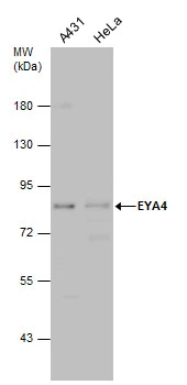
EYA4 antibodyGTX131408
ApplicationsWestern Blot
ReactivityHuman
TargetEYA4
- SizePrice
Product group Antibodies
EYA1/EYA4 AntibodyPACO07423
ApplicationsELISA, ImmunoHistoChemistry
ReactivityHuman, Mouse
TargetEYA4
- SizePrice