


Ezrin antibody [N2C2], Internal detects Ezrin protein on whole mount zebrafish by immunohistochemical analysis. Sample: Paraformaldehyde-fixed 2 days-post-fertilization zebrafish embryo. Green: Ezrin stained by Ezrin antibody [N2C2], Internal (GTX111709) diluted at 1:100. Antigen Retrieval: Tris-HCl buffer, pH 9.0, 20 min at 70oC
![Ezrin antibody [N2C2], Internal detects Ezrin protein on whole-mount zebrafish embryos by immunohistochemical analysis. Sample: Paraformaldehyde-fixed zebrafish embryos. Ezrin antibody [N2C2], Internal (GTX111709) dilution: 1:200.](https://www.genetex.com/upload/website/prouct_img/normal/GTX111709/GTX111709_40660_IHC-Wm_Z_22111423_954.webp "Ezrin antibody [N2C2], Internal detects Ezrin protein on whole-mount zebrafish embryos by immunohistochemical analysis. Sample: Paraformaldehyde-fixed zebrafish embryos. Ezrin antibody [N2C2], Internal (GTX111709) dilution: 1:200.")
![Ezrin antibody [N2C2], Internal detects Ezrin protein at cell membrane and cytoplasm by immunohistochemical analysis. Sample: Paraffin-embedded mouse tissue. Ezrin stained by Ezrin antibody [N2C2], Internal (GTX111709) diluted at 1:500. Antigen Retrieval: Citrate buffer, pH 6.0, 15 min](https://www.genetex.com/upload/website/prouct_img/normal/GTX111709/GTX111709_44461_20230414_IHC-P_multiple_M_23041023_437.webp "Ezrin antibody [N2C2], Internal detects Ezrin protein at cell membrane and cytoplasm by immunohistochemical analysis. Sample: Paraffin-embedded mouse tissue. Ezrin stained by Ezrin antibody [N2C2], Internal (GTX111709) diluted at 1:500. Antigen Retrieval: Citrate buffer, pH 6.0, 15 min")
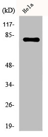
![Whole cell extract (30 μg) was separated by 7.5% SDS-PAGE, and the membrane was blotted with Ezrin antibody [N2C2], Internal (GTX111709) diluted at 1:1000. The HRP-conjugated anti-rabbit IgG antibody (GTX213110-01) was used to detect the primary antibody.](https://www.genetex.com/upload/website/prouct_img/normal/GTX111709/GTX111709_44461_20230414_WB_R_23041723_348.webp "Whole cell extract (30 μg) was separated by 7.5% SDS-PAGE, and the membrane was blotted with Ezrin antibody [N2C2], Internal (GTX111709) diluted at 1:1000. The HRP-conjugated anti-rabbit IgG antibody (GTX213110-01) was used to detect the primary antibody.")
![Whole cell extract (30 μg) was separated by 7.5% SDS-PAGE, and the membrane was blotted with Ezrin antibody [N2C2], Internal (GTX111709) diluted at 1:1000. The HRP-conjugated anti-rabbit IgG antibody (GTX213110-01) was used to detect the primary antibody.](https://www.genetex.com/upload/website/prouct_img/normal/GTX111709/GTX111709_44461_20230414_WB_M_23041723_827.webp "Whole cell extract (30 μg) was separated by 7.5% SDS-PAGE, and the membrane was blotted with Ezrin antibody [N2C2], Internal (GTX111709) diluted at 1:1000. The HRP-conjugated anti-rabbit IgG antibody (GTX213110-01) was used to detect the primary antibody.")
 antibody at 1:500 dilution.
Antigen Retrieval: Trilogy? (EDTA based, pH 8.0) buffer, 15min")
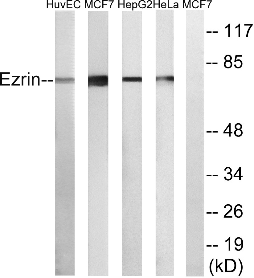
![Various whole cell extracts (30 μg) were separated by 7.5% SDS-PAGE, and the membrane was blotted with Ezrin antibody [N2C2], Internal (GTX111709) diluted at 1:10000. The HRP-conjugated anti-rabbit IgG antibody (GTX213110-01) was used to detect the primary antibody.](https://www.genetex.com/upload/website/prouct_img/normal/GTX111709/GTX111709_44461_20211008_WB_w_23060500_745.webp "Various whole cell extracts (30 μg) were separated by 7.5% SDS-PAGE, and the membrane was blotted with Ezrin antibody [N2C2], Internal (GTX111709) diluted at 1:10000. The HRP-conjugated anti-rabbit IgG antibody (GTX213110-01) was used to detect the primary antibody.")
![Ezrin antibody [N2C2], Internal detects Ezrin protein at cell membrane by immunofluorescent analysis. Sample: HeLa cells were fixed in 4% paraformaldehyde at RT for 15 min. Green: Ezrin stained by Ezrin antibody [N2C2], Internal (GTX111709) diluted at 1:500. Red: alpha Tubulin, a cytoskeleton marker, stained by alpha Tubulin antibody [GT114] (GTX628802) diluted at 1:1000. Blue: Fluoroshield with DAPI (GTX30920).](https://www.genetex.com/upload/website/prouct_img/normal/GTX111709/GTX111709_44482_20220121_ICC_IF_w_23060500_210.webp "Ezrin antibody [N2C2], Internal detects Ezrin protein at cell membrane by immunofluorescent analysis. Sample: HeLa cells were fixed in 4% paraformaldehyde at RT for 15 min. Green: Ezrin stained by Ezrin antibody [N2C2], Internal (GTX111709) diluted at 1:500. Red: alpha Tubulin, a cytoskeleton marker, stained by alpha Tubulin antibody [GT114] (GTX628802) diluted at 1:1000. Blue: Fluoroshield with DAPI (GTX30920).")
 antibody at 1:500 dilution.
Antigen Retrieval: Trilogy? (EDTA based, pH 8.0) buffer, 15min")
![Ezrin antibody [N2C2], Internal detects Ezrin protein at cytoplasm by immunofluorescent analysis. Sample: HCT116 cells were fixed in ice-cold MeOH for 5 min. Green: Ezrin protein stained by Ezrin antibody [N2C2], Internal (GTX111709) diluted at 1:1000. Blue: Hoechst 33342 staining.](https://www.genetex.com/upload/website/prouct_img/normal/GTX111709/GTX111709_40660_IFA_w_23060500_319.webp "Ezrin antibody [N2C2], Internal detects Ezrin protein at cytoplasm by immunofluorescent analysis. Sample: HCT116 cells were fixed in ice-cold MeOH for 5 min. Green: Ezrin protein stained by Ezrin antibody [N2C2], Internal (GTX111709) diluted at 1:1000. Blue: Hoechst 33342 staining.")
Ezrin antibody [N2C2], Internal detects Ezrin protein on whole mount zebrafish by immunohistochemical analysis. Sample: Paraformaldehyde-fixed 2 days-post-fertilization zebrafish embryo. Green: Ezrin stained by Ezrin antibody [N2C2], Internal (GTX111709) diluted at 1:100. Antigen Retrieval: Tris-HCl buffer, pH 9.0, 20 min at 70oC
Ezrin antibody [N2C2], Internal
GTX111709
ApplicationsImmunoFluorescence, Western Blot, ImmunoCytoChemistry, ImmunoHistoChemistry, ImmunoHistoChemistry Paraffin
Product group Antibodies
ReactivityHuman, Mouse, Rat, Zebra Fish
TargetEZR
Overview
- SupplierGeneTex
- Product NameEzrin antibody [N2C2], Internal
- Delivery Days Customer9
- Application Supplier NoteWB: 1:5000-1:20000. ICC/IF: 1:100-1:1000. IHC-P: 1:100-1:1000. *Optimal dilutions/concentrations should be determined by the researcher.Not tested in other applications.
- ApplicationsImmunoFluorescence, Western Blot, ImmunoCytoChemistry, ImmunoHistoChemistry, ImmunoHistoChemistry Paraffin
- CertificationResearch Use Only
- ClonalityPolyclonal
- Concentration1.84 mg/ml
- ConjugateUnconjugated
- Gene ID7430
- Target nameEZR
- Target descriptionezrin
- Target synonymsCVIL, CVL, HEL-S-105, VIL2, ezrin, cytovillin 2, epididymis secretory protein Li 105, p81, villin 2 (ezrin)
- HostRabbit
- IsotypeIgG
- Protein IDP15311
- Protein NameEzrin
- Scientific DescriptionThe cytoplasmic peripheral membrane protein encoded by this gene functions as a protein-tyrosine kinase substrate in microvilli. As a member of the ERM protein family, this protein serves as an intermediate between the plasma membrane and the actin cytoskeleton. This protein plays a key role in cell surface structure adhesion, migration and organization, and it has been implicated in various human cancers. A pseudogene located on chromosome 3 has been identified for this gene. Alternatively spliced variants have also been described for this gene. [provided by RefSeq]
- ReactivityHuman, Mouse, Rat, Zebra Fish
- Storage Instruction-20°C or -80°C,2°C to 8°C
- UNSPSC41116161
Datasheet
Related products
Product group Antibodies
Anti-Ezrin Antibody102-25737
ApplicationsWestern Blot, ImmunoHistoChemistry
TargetEZR
- SizePrice
Product group Antibodies
Anti-Ezrin AntibodyA95884
ApplicationsWestern Blot, ELISA, ImmunoHistoChemistry
ReactivityHuman, Mouse, Rat
- SizePrice
Product group Antibodies

Anti-EZR [SAIC-37C-23]Ab00330-1.1
ApplicationsMass Spectrometry, Western Blot
ReactivityHuman
TargetEZR
- SizePrice
Product group Antibodies

Ezrin (Phospho-Tyr146) AntibodyABX012576
ApplicationsWestern Blot, ELISA
- SizePrice

Product group Antibodies
Anti-Ezrin/EZR Antibody Picoband(r)A01750-2-CARRIER-FREE
ApplicationsFlow Cytometry, ImmunoFluorescence, Western Blot, ImmunoCytoChemistry, ImmunoHistoChemistry
ReactivityHuman, Monkey, Mouse, Rat
TargetEZR
- SizePrice

Product group Antibodies
Anti-EZR AntibodyAMAB90975
ApplicationsWestern Blot, ImmunoCytoChemistry, ImmunoHistoChemistry
ReactivityHuman
TargetEZR
- SizePrice
Product group Antibodies
Ezrin (EZR) Polyclonal AntibodyCAU26191
ApplicationsImmunoPrecipitation, Western Blot, ImmunoCytoChemistry, ImmunoHistoChemistry
ReactivityPorcine
TargetEZR
- SizePrice
Product group Antibodies
EZR AntibodyCSB-PA002456
ApplicationsWestern Blot, ELISA
ReactivityHuman, Mouse, Rat
TargetEZR
- SizePrice

Product group Antibodies
Ezrin (phospho Thr567) antibodyGTX133868
ApplicationsWestern Blot
ReactivityHuman
TargetEZR
- SizePrice
![IHC-P analysis of human lung tissue using GTX24069 Ezrin antibody [3C12].](https://www.genetex.com/upload/website/prouct_img/normal/GTX24069/GTX24069_20191203_IHC-P_111_w_23060722_704.webp)
Product group Antibodies
References
Ezrin antibody [3C12]GTX24069
ApplicationsImmunoHistoChemistry, ImmunoHistoChemistry Paraffin
ReactivityHuman, Mouse
TargetEZR
- SizePrice