

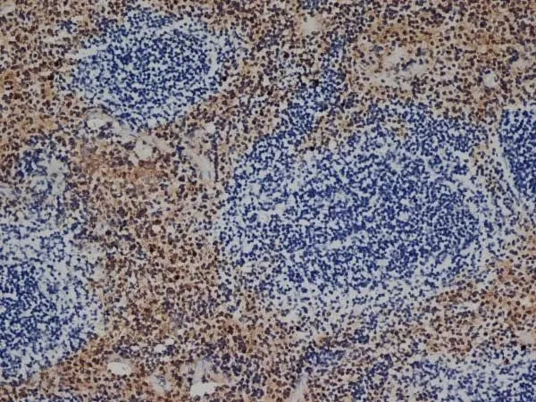
IHC-Fr analysis of mouse spleen tissue using GTX26640 F4/80 antibody [CI:A3-1].


IHC-Fr analysis of mouse spleen tissue using GTX26640 F4/80 antibody [CI:A3-1].
F4/80 antibody [CI:A3-1]
GTX26640
ApplicationsElectron Microscopy, Flow Cytometry, ImmunoFluorescence, ImmunoPrecipitation, Western Blot, ImmunoCytoChemistry, ImmunoHistoChemistry, ImmunoHistoChemistry Frozen, ImmunoHistoChemistry Paraffin, RadioImmunoAssay
Product group Antibodies
ReactivityHuman, Mouse, Rat
TargetAdgre1
Overview
- SupplierGeneTex
- Product NameF4/80 antibody [CI:A3-1]
- Delivery Days Customer9
- Application Supplier NoteFCM: 1/50-1/100. *Optimal dilutions/concentrations should be determined by the researcher.Not tested in other applications.
- ApplicationsElectron Microscopy, Flow Cytometry, ImmunoFluorescence, ImmunoPrecipitation, Western Blot, ImmunoCytoChemistry, ImmunoHistoChemistry, ImmunoHistoChemistry Frozen, ImmunoHistoChemistry Paraffin, RadioImmunoAssay
- CertificationResearch Use Only
- ClonalityMonoclonal
- Clone IDCI:A3-1
- Concentration1 mg/ml
- ConjugateUnconjugated
- Gene ID13733
- Target nameAdgre1
- Target descriptionadhesion G protein-coupled receptor E1
- Target synonymsDD7A5-7, EGF-TM7, Emr1, F4/80, Gpf480, Ly71, TM7LN3, adhesion G protein-coupled receptor E1, EGF-like module containing, mucin-like, hormone receptor-like sequence 1, EGF-like module receptor 1, EGF-like module-containing mucin-like hormone receptor-like 1, EMR1 hormone receptor, cell surface glycoprotein F4/80
- HostRat
- IsotypeIgG2b
- Protein IDQ61549
- Protein NameAdhesion G protein-coupled receptor E1
- ReactivityHuman, Mouse, Rat
- Storage Instruction-20°C or -80°C,2°C to 8°C
- UNSPSC41116161
References
- The potential role of hydrogen sulfide in regulating macrophage phenotypic changes via PINK1/parkin-mediated mitophagy in sepsis-related cardiorenal syndrome.Read this paper
- Adventitial macrophage accumulation impairs perivascular nerve function in mesenteric arteries with inflammatory bowel disease.Read this paper
- Sestrin2 Is Increased in Calcific Aortic Disease and Inhibits Osteoblastic Differentiation in Valvular Interstitial Cells via the Nuclear Factor E2-related Factor 2 Pathway.Read this paper
- Targeting prooxidant MnSOD effect inhibits triple-negative breast cancer (TNBC) progression and M2 macrophage functions under the oncogenic stress. Al Haq AT et al., 2022 Jan 11, Cell Death DisRead this paper
- Lupeol reduces M1 macrophage polarization to attenuate immunologic dissonance and fatty acid deposition in rats with diet-induced metabolic syndrome. Li J et al., 2021 Oct, Ann Transl MedRead this paper
Datasheet
Related products
Product group Antibodies

Anti-F4/80 [Cl:A3-1 (recombinant version)]AB00106-2.0-BT
ApplicationsFlow Cytometry, ImmunoHistoChemistry
ReactivityMouse
TargetAdgre1
- SizePrice

Product group Antibodies
Anti-F4/80/Adgre1 AntibodyA08751-CARRIER-FREE
ApplicationsImmunoFluorescence, ImmunoHistoChemistry
ReactivityMouse, Rat
TargetAdgre1
- SizePrice
Product group Antibodies
References
F4/80 antibody [CI:A3-1]GTX14321
ApplicationsFlow Cytometry, ImmunoFluorescence, Western Blot, ImmunoCytoChemistry, ImmunoHistoChemistry, ImmunoHistoChemistry Frozen, ImmunoHistoChemistry Paraffin, Other Application
ReactivityMouse
TargetAdgre1
- SizePrice
![FACS analysis of mouse C57Bl/6 bone marrow cells using GTX01470-02 F4/80 antibody [BM8.1] (Biotin). Right panel : co-stained with F4/80 antibody [BM8.1] (Biotin) and Mouse CD11b antibody (APC) Left panel : co-stained with isotype control and Mouse CD11b antibody (APC) antibody amount : 0.125 μg (5 μl)](https://www.genetex.com/upload/website/prouct_img/normal/GTX01470-02/GTX01470-02_20200428_FACS59_w_23053121_766.webp)
Product group Antibodies
F4/80 antibody [BM8.1] (Biotin)GTX01470-02
ApplicationsFlow Cytometry
ReactivityMouse
TargetAdgre1
- SizePrice
![FACS analysis of mouse C57Bl/6 bone marrow cells using GTX01470-06 F4/80 antibody [BM8.1] (FITC). Right panel : co-stained with F4/80 antibody [BM8.1] (FITC) and Mouse CD11b antibody (APC) Left panel : co-stained with isotype control and Mouse CD11b antibody (APC) antibody amount : 0.5 μg (5 μl)](https://www.genetex.com/upload/website/prouct_img/normal/GTX01470-06/GTX01470-06_20200428_FACS84_w_23053121_289.webp)
Product group Antibodies
F4/80 antibody [BM8.1] (FITC)GTX01470-06
ApplicationsFlow Cytometry
ReactivityMouse
TargetAdgre1
- SizePrice
![FACS analysis of mouse C57Bl/6 bone marrow cells using GTX01470-07 F4/80 antibody [BM8.1] (APC). Right panel : co-stained with F4/80 antibody [BM8.1] (APC) and Mouse CD11b antibody (FITC) Left panel : co-stained with isotype control and Mouse CD11b antibody (FITC) antibody amount : 0.5 μg (5 μl)](https://www.genetex.com/upload/website/prouct_img/normal/GTX01470-07/GTX01470-07_20200428_FACS26_w_23053121_791.webp)
Product group Antibodies
F4/80 antibody [BM8.1] (APC)GTX01470-07
ApplicationsFlow Cytometry
ReactivityMouse
TargetAdgre1
- SizePrice
![FACS analysis of mouse C57Bl/6 bone marrow cells using GTX01470-08 F4/80 antibody [BM8.1] (PE). Right panel : co-stained with F4/80 antibody [BM8.1] (PE) and Mouse CD11b antibody (APC) Left panel : co-stained with isotype control and Mouse CD11b antibody (APC) antibody amount : 0.25 μg (5 μl)](https://www.genetex.com/upload/website/prouct_img/normal/GTX01470-08/GTX01470-08_20200428_FACS119_w_23053121_411.webp)
Product group Antibodies
F4/80 antibody [BM8.1] (PE)GTX01470-08
ApplicationsFlow Cytometry
ReactivityMouse
TargetAdgre1
- SizePrice
![FACS analysis of mouse C57Bl/6 bone marrow cells using GTX01470-10 F4/80 antibody [BM8.1] (PE-Cy7). Right panel : co-stained with F4/80 antibody [BM8.1] (PE-Cy7) and Mouse CD11b antibody (APC) Left panel : co-stained with isotype control and Mouse CD11b antibody (APC) antibody amount : 0.25 μg (5 μl)](https://www.genetex.com/upload/website/prouct_img/normal/GTX01470-10/GTX01470-10_20200428_FACS148_w_23053121_677.webp)
Product group Antibodies
F4/80 antibody [BM8.1] (PE-Cy7)GTX01470-10
ApplicationsFlow Cytometry
ReactivityMouse
TargetAdgre1
- SizePrice
![FACS analysis of mouse C57Bl/6 bone marrow cells using GTX01470-11 F4/80 antibody [BM8.1] (PerCP-Cy5.5). Right panel : co-stained with F4/80 antibody [BM8.1] (PerCP-Cy5.5) and Mouse CD11b antibody (APC) Left panel : co-stained with isotype control and Mouse CD11b antibody (APC) antibody amount : 0.125 μg (5 μl)](https://www.genetex.com/upload/website/prouct_img/normal/GTX01470-11/GTX01470-11_20200428_FACS166_w_23053121_785.webp)
Product group Antibodies
F4/80 antibody [BM8.1] (PerCP-Cy5.5)GTX01470-11
ApplicationsFlow Cytometry
ReactivityMouse
TargetAdgre1
- SizePrice
![FACS analysis of mouse C57Bl/6 bone marrow cells using GTX01470-15 F4/80 antibody [BM8.1] (APC-Cy7). Right panel : co-stained with F4/80 antibody [BM8.1] (APC-Cy7) and Mouse CD11b antibody (violetFluor? 450) Left panel : co-stained with isotype control and Mouse CD11b antibody (violetFluor? 450) antibody amount : 0.25 μg (5 μl)](https://www.genetex.com/upload/website/prouct_img/normal/GTX01470-15/GTX01470-15_20200428_FACS48_w_23053121_853.webp)
Product group Antibodies
F4/80 antibody [BM8.1] (APC-Cy7)GTX01470-15
ApplicationsFlow Cytometry
ReactivityMouse
TargetAdgre1
- SizePrice