

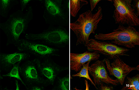
FACL4 antibody [C3], C-term detects FACL4 protein at cytoplasm by immunofluorescent analysis. Sample: HeLa cells were fixed in 4% paraformaldehyde at RT for 15 min. Green: FACL4 stained by FACL4 antibody [C3], C-term (GTX100260) diluted at 1:500. Red: alpha Tubulin, a cytoskeleton marker, stained by alpha Tubulin antibody [GT114] (GTX628802) diluted at 1:1000. Blue: Fluoroshield with DAPI (GTX30920).
![FACL4 antibody [C3], C-term detects FACL4 protein by immunohistochemical analysis. Sample: Paraffin-embedded rat tissues. FACL4 stained by FACL4 antibody [C3], C-term (GTX100260) diluted at 1:500. Antigen Retrieval: Citrate buffer, pH 6.0, 15 min](https://www.genetex.com/upload/website/prouct_img/normal/GTX100260/GTX100260_44832_20230414_IHC-P_multiple_R_23041719_873.webp "FACL4 antibody [C3], C-term detects FACL4 protein by immunohistochemical analysis. Sample: Paraffin-embedded rat tissues. FACL4 stained by FACL4 antibody [C3], C-term (GTX100260) diluted at 1:500. Antigen Retrieval: Citrate buffer, pH 6.0, 15 min")
![FACL4 antibody [C3], C-term detects FACL4 protein by immunohistochemical analysis. Sample: Paraffin-embedded mouse tissues. FACL4 stained by FACL4 antibody [C3], C-term (GTX100260) diluted at 1:500. Antigen Retrieval: Citrate buffer, pH 6.0, 15 min](https://www.genetex.com/upload/website/prouct_img/normal/GTX100260/GTX100260_44832_20230414_IHC-P_multiple_M_23041719_748.webp "FACL4 antibody [C3], C-term detects FACL4 protein by immunohistochemical analysis. Sample: Paraffin-embedded mouse tissues. FACL4 stained by FACL4 antibody [C3], C-term (GTX100260) diluted at 1:500. Antigen Retrieval: Citrate buffer, pH 6.0, 15 min")
![Immunoprecipitation of FACL4 protein from HeLa whole cell extracts using 5 μg of FACL4 antibody [C3], C-term (GTX100260). Western blot analysis was performed using FACL4 antibody [C3], C-term (GTX100260) diluted at 1:500. EasyBlot HRP-conjugated anti rabbit IgG antibody (GTX221666-01) was used to detect the primary antibody.](https://www.genetex.com/upload/website/prouct_img/normal/GTX100260/GTX100260_39568_20170629_IP_w_23060100_812.webp "Immunoprecipitation of FACL4 protein from HeLa whole cell extracts using 5 μg of FACL4 antibody [C3], C-term (GTX100260). Western blot analysis was performed using FACL4 antibody [C3], C-term (GTX100260) diluted at 1:500. EasyBlot HRP-conjugated anti rabbit IgG antibody (GTX221666-01) was used to detect the primary antibody.")
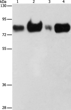
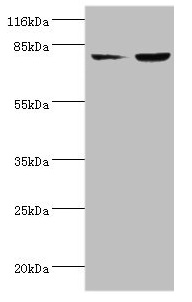
![Various whole cell extracts (30 μg) were separated by 7.5% SDS-PAGE, and the membrane was blotted with FACL4 antibody [C3], C-term (GTX100260) diluted at 1:1000. The HRP-conjugated anti-rabbit IgG antibody (GTX213110-01) was used to detect the primary antibody.](https://www.genetex.com/upload/website/prouct_img/normal/GTX100260/GTX100260_43831_20210611_WB_R_w_23060100_869.webp "Various whole cell extracts (30 μg) were separated by 7.5% SDS-PAGE, and the membrane was blotted with FACL4 antibody [C3], C-term (GTX100260) diluted at 1:1000. The HRP-conjugated anti-rabbit IgG antibody (GTX213110-01) was used to detect the primary antibody.")
![FACL4 antibody [C3], C-term detects FACL4 protein at cytosol on rat middle brain by immunohistochemical analysis. Sample: Paraffin-embedded rat middle brain. FACL4 antibody [C3], C-term (GTX100260) dilution: 1:500.
Antigen Retrieval: Trilogy? (EDTA based, pH 8.0) buffer, 15min](https://www.genetex.com/upload/website/prouct_img/normal/GTX100260/GTX100260_39568_IHC_R_w_23060100_300.webp "FACL4 antibody [C3], C-term detects FACL4 protein at cytosol on rat middle brain by immunohistochemical analysis. Sample: Paraffin-embedded rat middle brain. FACL4 antibody [C3], C-term (GTX100260) dilution: 1:500.
Antigen Retrieval: Trilogy? (EDTA based, pH 8.0) buffer, 15min")
![FACL4 antibody [C3], C-term detects FACL4 protein at cell membrane and cytoplasm by immunohistochemical analysis. Sample: Paraffin-embedded mouse kidney. FACL4 stained by FACL4 antibody [C3], C-term (GTX100260) diluted at 1:500. Antigen Retrieval: Citrate buffer, pH 6.0, 15 min](https://www.genetex.com/upload/website/prouct_img/normal/GTX100260/GTX100260_43831_20200724_IHC-P_M_w_23060100_444.webp "FACL4 antibody [C3], C-term detects FACL4 protein at cell membrane and cytoplasm by immunohistochemical analysis. Sample: Paraffin-embedded mouse kidney. FACL4 stained by FACL4 antibody [C3], C-term (GTX100260) diluted at 1:500. Antigen Retrieval: Citrate buffer, pH 6.0, 15 min")
![Various whole cell extracts (30 μg) were separated by 7.5% SDS-PAGE, and the membrane was blotted with FACL4 antibody [C3], C-term (GTX100260) diluted at 1:1000. The HRP-conjugated anti-rabbit IgG antibody (GTX213110-01) was used to detect the primary antibody.](https://www.genetex.com/upload/website/prouct_img/normal/GTX100260/GTX100260_43831_20210611_WB_M_w_23060100_912.webp "Various whole cell extracts (30 μg) were separated by 7.5% SDS-PAGE, and the membrane was blotted with FACL4 antibody [C3], C-term (GTX100260) diluted at 1:1000. The HRP-conjugated anti-rabbit IgG antibody (GTX213110-01) was used to detect the primary antibody.")
![FACL4 antibody [C3], C-term detects FACL4 protein at cytosol on mouse lymph node by immunohistochemical analysis. Sample: Paraffin-embedded mouse lymph node. FACL4 antibody [C3], C-term (GTX100260) dilution: 1:500.
Antigen Retrieval: Trilogy? (EDTA based, pH 8.0) buffer, 15min](https://www.genetex.com/upload/website/prouct_img/normal/GTX100260/GTX100260_39568_IHC_M_w_23060100_235.webp "FACL4 antibody [C3], C-term detects FACL4 protein at cytosol on mouse lymph node by immunohistochemical analysis. Sample: Paraffin-embedded mouse lymph node. FACL4 antibody [C3], C-term (GTX100260) dilution: 1:500.
Antigen Retrieval: Trilogy? (EDTA based, pH 8.0) buffer, 15min")
![Various whole cell extracts (30 μg) were separated by 7.5% SDS-PAGE, and the membrane was blotted with FACL4 antibody [C3], C-term (GTX100260) diluted at 1:1000. The HRP-conjugated anti-rabbit IgG antibody (GTX213110-01) was used to detect the primary antibody.](https://www.genetex.com/upload/website/prouct_img/normal/GTX100260/GTX100260_44832_20221014_WB_24112622_246.webp "Various whole cell extracts (30 μg) were separated by 7.5% SDS-PAGE, and the membrane was blotted with FACL4 antibody [C3], C-term (GTX100260) diluted at 1:1000. The HRP-conjugated anti-rabbit IgG antibody (GTX213110-01) was used to detect the primary antibody.")
FACL4 antibody [C3], C-term detects FACL4 protein at cytoplasm by immunofluorescent analysis. Sample: HeLa cells were fixed in 4% paraformaldehyde at RT for 15 min. Green: FACL4 stained by FACL4 antibody [C3], C-term (GTX100260) diluted at 1:500. Red: alpha Tubulin, a cytoskeleton marker, stained by alpha Tubulin antibody [GT114] (GTX628802) diluted at 1:1000. Blue: Fluoroshield with DAPI (GTX30920).
FACL4 antibody [C3], C-term
GTX100260
ApplicationsImmunoFluorescence, ImmunoPrecipitation, Western Blot, ImmunoCytoChemistry, ImmunoHistoChemistry, ImmunoHistoChemistry Paraffin
Product group Antibodies
ReactivityHuman, Mouse, Rat
TargetACSL4
Overview
- SupplierGeneTex
- Product NameFACL4 antibody [C3], C-term
- Delivery Days Customer9
- Application Supplier NoteWB: 1:500-1:10000. ICC/IF: 1:100-1:1000. IHC-P: 1:100-1:1000. IP: 1:1000-1:5000. *Optimal dilutions/concentrations should be determined by the researcher.Not tested in other applications.
- ApplicationsImmunoFluorescence, ImmunoPrecipitation, Western Blot, ImmunoCytoChemistry, ImmunoHistoChemistry, ImmunoHistoChemistry Paraffin
- CertificationResearch Use Only
- ClonalityPolyclonal
- Concentration1.37 mg/ml
- ConjugateUnconjugated
- Gene ID2182
- Target nameACSL4
- Target descriptionacyl-CoA synthetase long chain family member 4
- Target synonymsACS4, FACL4, LACS4, MRX63, MRX68, XLID63, long-chain-fatty-acid--CoA ligase 4, acyl-CoA synthetase 4, arachidonate--CoA ligase, fatty-acid-Coenzyme A ligase, long-chain 4, lignoceroyl-CoA synthase, long-chain acyl-CoA synthetase 4, long-chain fatty-acid-Coenzyme A ligase 4
- HostRabbit
- IsotypeIgG
- Protein IDO60488
- Protein NameLong-chain-fatty-acid--CoA ligase 4
- Scientific DescriptionThe protein encoded by this gene is an isozyme of the long-chain fatty-acid-coenzyme A ligase family. Although differing in substrate specificity, subcellular localization, and tissue distribution, all isozymes of this family convert free long-chain fatty acids into fatty acyl-CoA esters, and thereby play a key role in lipid biosynthesis and fatty acid degradation. This isozyme preferentially utilizes arachidonate as substrate. The absence of this enzyme may contribute to the mental retardation or Alport syndrome. Alternative splicing of this gene generates 2 transcript variants. [provided by RefSeq]
- ReactivityHuman, Mouse, Rat
- Storage Instruction-20°C or -80°C,2°C to 8°C
- UNSPSC41116161
Datasheet
Related products
Product group Antibodies
Anti-ACSL4 AntibodyA37874
ApplicationsWestern Blot, ImmunoHistoChemistry
ReactivityHuman
- SizePrice

Product group Antibodies
Anti-FACL4/ACSL4 Antibody Picoband(r)A04372-2-CARRIER-FREE
ApplicationsFlow Cytometry, ImmunoFluorescence, Western Blot, ImmunoCytoChemistry, ImmunoHistoChemistry
ReactivityHuman, Mouse, Rat
TargetACSL4
- SizePrice
Product group Antibodies
Anti-ACSL4 Antibody144-06826
ApplicationsWestern Blot
ReactivityHuman, Mouse
TargetACSL4
- SizePrice
Product group Antibodies
ACSL4 Recombinant AntibodyBSM-62432R
ApplicationsImmunoFluorescence, ImmunoPrecipitation, Western Blot, ImmunoHistoChemistry, ImmunoHistoChemistry Frozen, ImmunoHistoChemistry Paraffin
ReactivityHuman, Mouse, Rat
TargetACSL4
- SizePrice
Product group Antibodies
References

Goat anti-FACL4 / ACSL4EB05662
ApplicationsFlow Cytometry, ImmunoFluorescence, Western Blot, ELISA, ImmunoHistoChemistry
ReactivityBovine, Canine, Human, Mouse, Porcine, Rat
TargetACSL4
- SizePrice
Product group Antibodies
Acsl4 Polyclonal AntibodyCAC09075
ApplicationsWestern Blot, ELISA, ImmunoHistoChemistry
TargetACSL4
- SizePrice
Product group Antibodies
ACSL4 AntibodyCSB-PA060488EA01HU
ApplicationsWestern Blot, ELISA, ImmunoHistoChemistry
ReactivityHuman
TargetACSL4
- SizePrice
Product group Antibodies

ACSL4 / FACL4 AntibodyLS-C404608
ApplicationsWestern Blot, ELISA, ImmunoHistoChemistry
ReactivityHuman, Mouse, Rat
TargetACSL4
- SizePrice
Product group Antibodies
FACL4 antibody, C-termGTX89766
ApplicationsWestern Blot, ImmunoHistoChemistry, ImmunoHistoChemistry Paraffin
ReactivityHuman
TargetACSL4
- SizePrice