

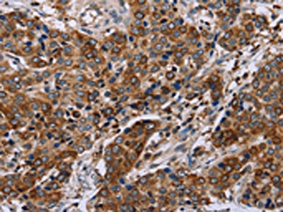
The image on the left is immunohistochemistry of paraffin-embedded Human liver cancer tissue using CSB-PA993404(FAF1 Antibody) at dilution 1/50, on the right is treated with synthetic peptide. (Original magnification: x200)
 at dilution 1/50, on the right is treated with synthetic peptide. (Original magnification: x200)")
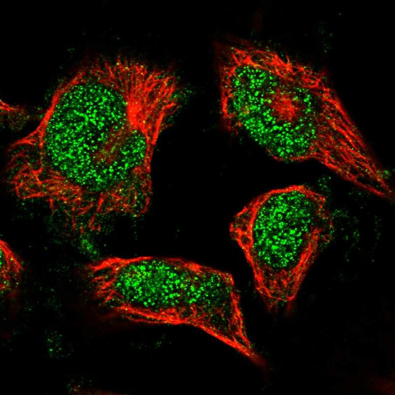
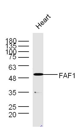
 at dilution 1/400, Secondary antibody: Goat anti rabbit IgG at 1/8000 dilution, Exposure time: 5 minutes")
The image on the left is immunohistochemistry of paraffin-embedded Human liver cancer tissue using CSB-PA993404(FAF1 Antibody) at dilution 1/50, on the right is treated with synthetic peptide. (Original magnification: x200)
FAF1 Antibody
CSB-PA993404
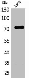
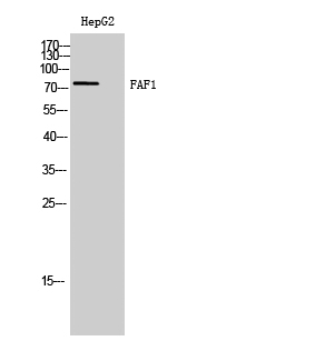
ApplicationsWestern Blot, ELISA, ImmunoHistoChemistry
Product group Antibodies
TargetFAF1
Overview
- SupplierCusabio
- Product NameFAF1 Antibody
- Delivery Days Customer20
- ApplicationsWestern Blot, ELISA, ImmunoHistoChemistry
- CertificationResearch Use Only
- ClonalityPolyclonal
- ConjugateUnconjugated
- Gene ID11124
- Target nameFAF1
- Target descriptionFas associated factor 1
- Target synonymsCGI-03; Fas (TNFRSF6) associated factor 1; FAS-associated factor 1; hFAF1; HFAF1s; TNFRSF6-associated factor 1; UBX domain protein 3A; UBX domain-containing protein 12; UBX domain-containing protein 3A; UBXD12; UBXN3A
- HostRabbit
- IsotypeIgG
- Protein IDQ9UNN5
- Protein NameFAS-associated factor 1
- Scientific DescriptionInteraction of Fas ligand (TNFSF6) with the FAS antigen (TNFRSF6) mediates programmed cell death, also called apoptosis, in a number of organ systems. The protein encoded by this gene binds to FAS antigen and can initiate apoptosis or enhance apoptosis initiated through FAS antigen. Initiation of apoptosis by the protein encoded by this gene requires a ubiquitin-like domain but not the FAS-binding domain.
- Storage Instruction-20°C or -80°C
- UNSPSC12352203
Related products
Product group Antibodies
FAF1 Polyclonal AntibodyCAC14672
ApplicationsWestern Blot, ELISA, ImmunoHistoChemistry
TargetFAF1
- SizePrice
Product group Antibodies
Anti-FAF1 AntibodyHPA018253
ApplicationsImmunoCytoChemistry
TargetFAF1
- SizePrice
Product group Antibodies
ApplicationsImmunoPrecipitation, Western Blot, ImmunoHistoChemistry
TargetFAF1
- SizePrice

Product group Antibodies
Anti-FAF1 Antibody Picoband(r)A03842-1-CARRIER-FREE
ApplicationsFlow Cytometry, ImmunoFluorescence, Western Blot, ELISA, ImmunoCytoChemistry
TargetFAF1
- SizePrice
Product group Antibodies
FAF1 Polyclonal AntibodyBS-1348R
ApplicationsFlow Cytometry, ImmunoFluorescence, Western Blot, ELISA, ImmunoCytoChemistry, ImmunoHistoChemistry, ImmunoHistoChemistry Frozen, ImmunoHistoChemistry Paraffin
TargetFAF1
- SizePrice