

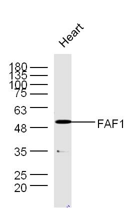
Mouse heart lysates probed with Rabbit Anti-FAF1 Polyclonal Antibody, Unconjugated (bs-1348R) at 1:300 overnight at 4˚C. Followed by a conjugated secondary antibody for 90 min at 37˚C.
 followed by conjugation to the secondary antibody and DAB staining")
; Antigen retrieval by boiling in sodium citrate buffer (pH6.0) for 15min; Block endogenous peroxidase by 3% hydrogen peroxide for 20 minutes; Blocking buffer (normal goat serum) at 37°C for 30min; Antibody incubation with (FAF1) Polyclonal Antibody, Unconjugated (bs-1348R) at 1:200 overnight at 4°C, followed by operating according to SP Kit(Rabbit) (sp-0023) instructionsand DAB staining.")
Mouse heart lysates probed with Rabbit Anti-FAF1 Polyclonal Antibody, Unconjugated (bs-1348R) at 1:300 overnight at 4˚C. Followed by a conjugated secondary antibody for 90 min at 37˚C.
FAF1 Polyclonal Antibody
BS-1348R
ApplicationsFlow Cytometry, ImmunoFluorescence, Western Blot, ELISA, ImmunoCytoChemistry, ImmunoHistoChemistry, ImmunoHistoChemistry Frozen, ImmunoHistoChemistry Paraffin
Product group Antibodies
ReactivityBovine, Canine, Chicken, Human, Mouse, Rabbit, Rat
TargetFAF1
Overview
- SupplierBioss
- Product NameFAF1 Polyclonal Antibody
- Delivery Days Customer16
- ApplicationsFlow Cytometry, ImmunoFluorescence, Western Blot, ELISA, ImmunoCytoChemistry, ImmunoHistoChemistry, ImmunoHistoChemistry Frozen, ImmunoHistoChemistry Paraffin
- Applications SupplierWB(1:300-5000), ELISA(1:500-1000), FCM(1:20-100), IHC-P(1:200-400), IHC-F(1:100-500), IF(IHC-P)(1:50-200), IF(IHC-F)(1:50-200), IF(ICC)(1:50-200)
- CertificationResearch Use Only
- ClonalityPolyclonal
- Concentration1 ug/ul
- ConjugateUnconjugated
- Gene ID11124
- Target nameFAF1
- Target descriptionFas associated factor 1
- Target synonymsCGI-03, HFAF1s, UBXD12, UBXN3A, hFAF1, FAS-associated factor 1, Fas (TNFRSF6) associated factor 1, TNFRSF6-associated factor 1, UBX domain protein 3A, UBX domain-containing protein 12, UBX domain-containing protein 3A
- HostRabbit
- IsotypeIgG
- Protein IDQ9UNN5
- Protein NameFAS-associated factor 1
- ReactivityBovine, Canine, Chicken, Human, Mouse, Rabbit, Rat
- Storage Instruction-20°C
- UNSPSC41116161
Datasheet
Related products
Product group Antibodies
Anti-FAF1 AntibodyA98159
ApplicationsWestern Blot, ELISA
ReactivityHuman, Mouse, Rat
- SizePrice

Product group Antibodies
Anti-FAF1 Antibody Picoband(r)A03842-1-CARRIER-FREE
ApplicationsFlow Cytometry, ImmunoFluorescence, Western Blot, ELISA, ImmunoCytoChemistry
ReactivityHuman, Mouse, Rat
TargetFAF1
- SizePrice
Product group Antibodies
Anti-FAF1 Antibody144-61354
ApplicationsWestern Blot
ReactivityHuman, Mouse, Rat
TargetFAF1
- SizePrice
Product group Antibodies
FAF1 Polyclonal AntibodyCAC14672
ApplicationsWestern Blot, ELISA, ImmunoHistoChemistry
ReactivityMouse
TargetFAF1
- SizePrice
Product group Antibodies
FAF1 AntibodyCSB-PA004921
ApplicationsWestern Blot, ELISA
ReactivityHuman, Mouse, Rat
TargetFAF1
- SizePrice
Product group Antibodies
FAF1 antibodyGTX102369
ApplicationsWestern Blot
ReactivityHuman, Mouse
TargetFAF1
- SizePrice
Product group Antibodies
Anti-FAF1 AntibodyHPA018253
ApplicationsImmunoCytoChemistry
ReactivityHuman
TargetFAF1
- SizePrice
Product group Antibodies
Anti-FAF1 AntibodyCAB2921
ApplicationsWestern Blot, ELISA
ReactivityHuman
TargetFAF1
- SizePrice