

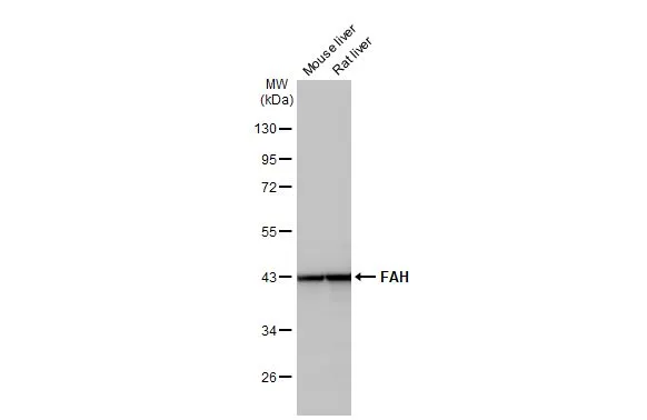
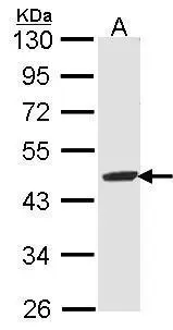
Various tissue extracts (50 μg) were separated by 10% SDS-PAGE, and the membrane was blotted with FAH antibody [HL1972] (GTX637865) diluted at 1:1000. The HRP-conjugated anti-rabbit IgG antibody (GTX213110-01) was used to detect the primary antibody.
![Various tissue extracts (50 μg) were separated by 10% SDS-PAGE, and the membrane was blotted with FAH antibody [HL1972] (GTX637865) diluted at 1:1000. The HRP-conjugated anti-rabbit IgG antibody (GTX213110-01) was used to detect the primary antibody.](https://www.genetex.com/upload/website/prouct_img/normal/GTX637865/GTX637865_T-44858_20221125_WB_M_tissue_22112723_174.webp "Various tissue extracts (50 μg) were separated by 10% SDS-PAGE, and the membrane was blotted with FAH antibody [HL1972] (GTX637865) diluted at 1:1000. The HRP-conjugated anti-rabbit IgG antibody (GTX213110-01) was used to detect the primary antibody.")
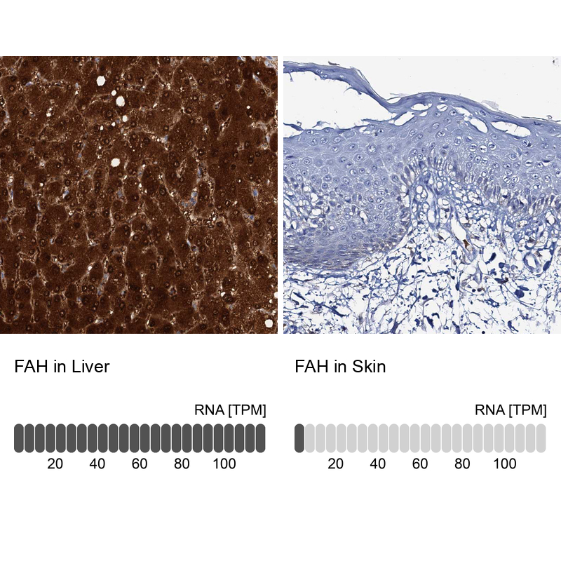
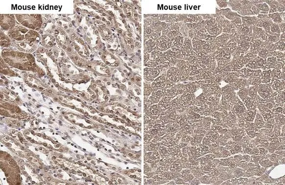
![FAH antibody [HL1972] detects FAH protein at cytoplasm by immunohistochemical analysis. Sample: Paraffin-embedded mouse liver. FAH stained by FAH antibody [HL1972] (GTX637865) diluted at 1:100. Antigen Retrieval: Citrate buffer, pH 6.0, 15 min](https://www.genetex.com/upload/website/prouct_img/normal/GTX637865/GTX637865_T-44858_20221111_IHC-P_M_22122018_189.webp "FAH antibody [HL1972] detects FAH protein at cytoplasm by immunohistochemical analysis. Sample: Paraffin-embedded mouse liver. FAH stained by FAH antibody [HL1972] (GTX637865) diluted at 1:100. Antigen Retrieval: Citrate buffer, pH 6.0, 15 min")
![FAH antibody [HL1972] detects FAH protein at cytoplasm by immunohistochemical analysis. Sample: Paraffin-embedded rat liver. FAH stained by FAH antibody [HL1972] (GTX637865) diluted at 1:100. Antigen Retrieval: Citrate buffer, pH 6.0, 15 min](https://www.genetex.com/upload/website/prouct_img/normal/GTX637865/GTX637865_T-44858_20221111_IHC-P_R_22122018_557.webp "FAH antibody [HL1972] detects FAH protein at cytoplasm by immunohistochemical analysis. Sample: Paraffin-embedded rat liver. FAH stained by FAH antibody [HL1972] (GTX637865) diluted at 1:100. Antigen Retrieval: Citrate buffer, pH 6.0, 15 min")
![Various whole cell extracts (30 μg) were separated by 10% SDS-PAGE, and the membrane was blotted with FAH antibody [HL1972] (GTX637865) diluted at 1:1000. The HRP-conjugated anti-rabbit IgG antibody (GTX213110-01) was used to detect the primary antibody. Corresponding RNA expression data for the same cell lines are based on Human Protein Atlas program.](https://www.genetex.com/upload/website/prouct_img/normal/GTX637865/GTX637865_44907_20221230_WB_TPM_watermark_23010400_746.webp "Various whole cell extracts (30 μg) were separated by 10% SDS-PAGE, and the membrane was blotted with FAH antibody [HL1972] (GTX637865) diluted at 1:1000. The HRP-conjugated anti-rabbit IgG antibody (GTX213110-01) was used to detect the primary antibody. Corresponding RNA expression data for the same cell lines are based on Human Protein Atlas program.")
![Non-transfected (–) and transfected (+) 293T whole cell extracts (30 μg) were separated by 10% SDS-PAGE, and the membrane was blotted with FAH antibody [HL1972] (GTX637865) diluted at 1:1000. The HRP-conjugated anti-rabbit IgG antibody (GTX213110-01) was used to detect the primary antibody.](https://www.genetex.com/upload/website/prouct_img/normal/GTX637865/GTX637865_44907_20230217_WB_shRNA_watermark_23022022_647.webp "Non-transfected (–) and transfected (+) 293T whole cell extracts (30 μg) were separated by 10% SDS-PAGE, and the membrane was blotted with FAH antibody [HL1972] (GTX637865) diluted at 1:1000. The HRP-conjugated anti-rabbit IgG antibody (GTX213110-01) was used to detect the primary antibody.")
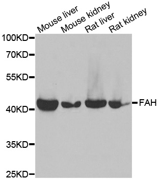
Various tissue extracts (50 μg) were separated by 10% SDS-PAGE, and the membrane was blotted with FAH antibody [HL1972] (GTX637865) diluted at 1:1000. The HRP-conjugated anti-rabbit IgG antibody (GTX213110-01) was used to detect the primary antibody.
FAH antibody [HL1972]
GTX637865
ApplicationsWestern Blot, ImmunoHistoChemistry, ImmunoHistoChemistry Paraffin
Product group Antibodies
ReactivityHuman, Mouse, Rat
TargetFAH
Overview
- SupplierGeneTex
- Product NameFAH antibody [HL1972]
- Delivery Days Customer9
- Application Supplier NoteWB: 1:500-1:3000. *Optimal dilutions/concentrations should be determined by the researcher.Not tested in other applications.
- ApplicationsWestern Blot, ImmunoHistoChemistry, ImmunoHistoChemistry Paraffin
- CertificationResearch Use Only
- ClonalityMonoclonal
- Clone IDHL1972
- Concentration1 mg/ml
- ConjugateUnconjugated
- Gene ID2184
- Target nameFAH
- Target descriptionfumarylacetoacetate hydrolase
- Target synonymsfumarylacetoacetase, FAA, beta-diketonase, epididymis secretory sperm binding protein, fumarylacetoacetate hydrolase (fumarylacetoacetase)
- HostRabbit
- IsotypeIgG
- Protein IDP16930
- Protein NameFumarylacetoacetase
- Scientific DescriptionThis gene encodes the last enzyme in the tyrosine catabolism pathway. FAH deficiency is associated with Type 1 hereditary tyrosinemia (HT). [provided by RefSeq, Jul 2008]
- ReactivityHuman, Mouse, Rat
- Storage Instruction-20°C or -80°C,2°C to 8°C
- UNSPSC41116161
Datasheet
Related products
Product group Antibodies
FAH AntibodyCSB-PA007965LA01HU
ApplicationsImmunoFluorescence, Western Blot, ELISA, ImmunoHistoChemistry
ReactivityHuman, Mouse
TargetFAH
- SizePrice

Product group Antibodies
Anti-FAA/FAH Antibody Picoband(r)A02072-1-CARRIER-FREE
ApplicationsFlow Cytometry, Western Blot, ELISA, ImmunoHistoChemistry
ReactivityHuman, Mouse, Rat
TargetFAH
- SizePrice
Product group Antibodies
Anti-FAH AntibodyA31436
ApplicationsWestern Blot, ImmunoHistoChemistry
ReactivityHuman, Mouse, Rat
- SizePrice
Product group Antibodies

FAH AntibodyLS-C832017
ApplicationsWestern Blot, ELISA, ImmunoHistoChemistry
ReactivityHuman, Mouse, Rat
TargetFAH
- SizePrice
Product group Antibodies
Anti-FAH AntibodyHPA041370
ApplicationsWestern Blot, ImmunoCytoChemistry, ImmunoHistoChemistry
ReactivityHuman
TargetFAH
- SizePrice
Product group Antibodies
Fah Polyclonal AntibodyCAC08422
ApplicationsImmunoFluorescence, Western Blot, ELISA, ImmunoHistoChemistry
ReactivityMouse
TargetFAH
- SizePrice
![Various tissue extracts (50 μg) were separated by 10% SDS-PAGE, and the membrane was blotted with FAH antibody [HL1970] (GTX637863) diluted at 1:1000. The HRP-conjugated anti-rabbit IgG antibody (GTX213110-01) was used to detect the primary antibody.](https://www.genetex.com/upload/website/prouct_img/normal/GTX637863/GTX637863_T-44858_20221125_WB_M_tissue_22112723_506.webp)
Product group Antibodies
FAH antibody [HL1970]GTX637863
ApplicationsWestern Blot, ImmunoHistoChemistry, ImmunoHistoChemistry Paraffin
ReactivityHuman, Mouse, Rat
TargetFAH
- SizePrice
Product group Antibodies
FAH antibodyGTX102583
ApplicationsWestern Blot
ReactivityHuman
TargetFAH
- SizePrice
Product group Antibodies
FAH antibodyGTX114400
ApplicationsImmunoFluorescence, Western Blot, ImmunoCytoChemistry, ImmunoHistoChemistry, ImmunoHistoChemistry Frozen, ImmunoHistoChemistry Paraffin
ReactivityHuman, Mouse, Rat
TargetFAH
- SizePrice