

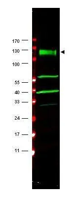
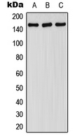
Western blot using GeneTex's affinity purified anti-FANCA antibody shows detection of a band at ~133 kDa (arrowhead) corres-ponding to FANCA in HeLa whole cell lysates. The identity of the lower molecular weight bands is unknown but may represent breakdown products. Approximately 35 ug of lysate was separated by 4-20% Tris Glycine SDS-PAGE. After blocking, the membrane was probed for 2 h at room temperature with the primary antibody diluted to 1:1,500. The membrane was washed and reacted with a 1:10,000 dilution of IRDye?800 conjugated Gt-a-Rabbit IgG [H&L] for 45 min at room temperature (800 nm channel, green). Molecular weight estimation was made by comparison to prestained MW markers indicated at left (700 nm channel, red). IRDye?800 fluorescence images were captured using the OdysseyR Infrared Imaging System developed by LI-COR. IRDye is a trademark of LI-COR, Inc. Other detection systems will yield similar results.

Western blot using GeneTex's affinity purified anti-FANCA antibody shows detection of a band at ~133 kDa (arrowhead) corres-ponding to FANCA in HeLa whole cell lysates. The identity of the lower molecular weight bands is unknown but may represent breakdown products. Approximately 35 ug of lysate was separated by 4-20% Tris Glycine SDS-PAGE. After blocking, the membrane was probed for 2 h at room temperature with the primary antibody diluted to 1:1,500. The membrane was washed and reacted with a 1:10,000 dilution of IRDye?800 conjugated Gt-a-Rabbit IgG [H&L] for 45 min at room temperature (800 nm channel, green). Molecular weight estimation was made by comparison to prestained MW markers indicated at left (700 nm channel, red). IRDye?800 fluorescence images were captured using the OdysseyR Infrared Imaging System developed by LI-COR. IRDye is a trademark of LI-COR, Inc. Other detection systems will yield similar results.
FANCA antibody
GTX25063
ApplicationsWestern Blot, ELISA
Product group Antibodies
ReactivityHuman
TargetFANCA
Overview
- SupplierGeneTex
- Product NameFANCA antibody
- Delivery Days Customer9
- Application Supplier NoteWB: 1:500-1:3000. ELISA: 1:15000-1:30000. *Optimal dilutions/concentrations should be determined by the researcher.Not tested in other applications.
- ApplicationsWestern Blot, ELISA
- CertificationResearch Use Only
- ClonalityPolyclonal
- Concentration2.2 mg/ml
- ConjugateUnconjugated
- Gene ID2175
- Target nameFANCA
- Target descriptionFA complementation group A
- Target synonymsFA, FA-H, FA1, FAA, FACA, FAH, FANCH, Fanconi anemia group A protein, Fanconi anemia complementation group A, Fanconi anemia, complementation group H, Fanconi anemia, type 1
- HostRabbit
- IsotypeIgG
- Protein IDO15360
- Protein NameFanconi anemia group A protein
- Scientific DescriptionFANCA is a DNA repair protein that may operate in a postreplication repair or a cell cycle checkpoint function. It may be involved in interstrand DNA cross-link repair and in the maintenance of normal chromosome stability.
- ReactivityHuman
- Storage Instruction-20°C or -80°C,2°C to 8°C
- UNSPSC41116161
Datasheet
Related products
Product group Antibodies
Anti-FANCA Antibody144-07671
ApplicationsWestern Blot, ImmunoHistoChemistry
ReactivityHuman
TargetFANCA
- SizePrice
Product group Antibodies
Anti-FANCA AntibodyA100674
ApplicationsELISA, ImmunoHistoChemistry
ReactivityHuman
- SizePrice
Product group Antibodies
FANCA Polyclonal AntibodyCAC13035
ApplicationsImmunoFluorescence, ELISA
TargetFANCA
- SizePrice
Product group Antibodies
Phospho-FANCA (S1149) AntibodyCSB-PA008175
ApplicationsImmunoFluorescence, Western Blot, ELISA, ImmunoHistoChemistry
ReactivityHuman
TargetFANCA
- SizePrice
Product group Antibodies
FANCA Polyclonal AntibodyBS-13138R
ApplicationsImmunoFluorescence, ELISA, ImmunoCytoChemistry, ImmunoHistoChemistry, ImmunoHistoChemistry Frozen, ImmunoHistoChemistry Paraffin
ReactivityChicken, Human, Mouse, Rabbit, Rat
TargetFANCA
- SizePrice
Product group Antibodies
FANCA antibody [N1], N-termGTX113433
ApplicationsImmunoFluorescence, Western Blot, ImmunoCytoChemistry, ImmunoHistoChemistry, ImmunoHistoChemistry Paraffin
ReactivityHuman
TargetFANCA
- SizePrice
Product group Antibodies
FANCA (phospho Ser1149) antibodyGTX32179
ApplicationsImmunoFluorescence, Western Blot, ImmunoCytoChemistry
ReactivityHuman, Mouse, Rat
TargetFANCA
- SizePrice

Product group Antibodies
Anti-FANCA AntibodyHPA063236
ApplicationsImmunoCytoChemistry
ReactivityHuman
TargetFANCA
- SizePrice
Product group Antibodies

FANCA AntibodyLS-C409225
ApplicationsWestern Blot, ImmunoHistoChemistry
ReactivityHuman
TargetFANCA
- SizePrice