

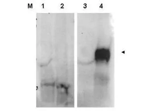
Western blot using GeneTex's affinity purified anti-FAP antibody shows detection of FAP protein in whole cell lysates from FAP expressing HEK cells (lane 4) but not control HEK cells (lane 3). Specific band staining is blocked when the primary antibody is pre-incubated with immunizing peptide (lanes 1 and 2 respectively). The band at ~90 kDa, indicated by the arrowhead, corresponds to the expected molecular weight of transfected FAP. Primary antibody was used at 1:1,000.

Western blot using GeneTex's affinity purified anti-FAP antibody shows detection of FAP protein in whole cell lysates from FAP expressing HEK cells (lane 4) but not control HEK cells (lane 3). Specific band staining is blocked when the primary antibody is pre-incubated with immunizing peptide (lanes 1 and 2 respectively). The band at ~90 kDa, indicated by the arrowhead, corresponds to the expected molecular weight of transfected FAP. Primary antibody was used at 1:1,000.
FAP antibody
GTX48711
ApplicationsWestern Blot, ELISA
Product group Antibodies
ReactivityMouse
TargetFap
Overview
- SupplierGeneTex
- Product NameFAP antibody
- Delivery Days Customer9
- Application Supplier NoteWB: 1:500-1:2000. ELISA: 1:10000-1:20000. *Optimal dilutions/concentrations should be determined by the researcher.Not tested in other applications.
- ApplicationsWestern Blot, ELISA
- CertificationResearch Use Only
- ClonalityPolyclonal
- Concentration1.1 mg/ml
- ConjugateUnconjugated
- Gene ID14089
- Target nameFap
- Target descriptionfibroblast activation protein
- Target synonymsSIMP, prolyl endopeptidase FAP, FAPalpha, dipeptidyl peptidase FAP, fibroblast activation protein alpha, gelatine degradation protease FAP, integral membrane serine protease, post-proline cleaving enzyme, seprase, serine integral membrane protease, surface-expressed protease
- HostRabbit
- IsotypeIgG
- Protein IDP97321
- Protein NameProlyl endopeptidase FAP
- Scientific DescriptionFibroblast Activation Protein (FAP) is expressed in the stroma of sites that are undergoing wound healing. In addition, it has recently been reported that FAP is expressed in the stroma of sites of metastatic disease. Inhibition of FAP may lead to a dramatic decrease in the number of metastatic osteosarcoma lung nodules. FAP exists as an inactive monomer and when activated forms homodimers or heterodimers with DPP4. Multiple isoforms of FAP are reported as alternative splicing products from a common gene.
- ReactivityMouse
- Storage Instruction-20°C or -80°C,2°C to 8°C
- UNSPSC41116161
Datasheet
Related products
Product group Antibodies

Anti-FAP [73.3]Ab03229-1.1
ApplicationsFlow Cytometry, ImmunoPrecipitation, Western Blot, Other Application
ReactivityMouse
TargetFap
- SizePrice
Product group Antibodies
ApplicationsImmunoPrecipitation, Western Blot, ImmunoCytoChemistry, ImmunoHistoChemistry
ReactivityMouse
TargetFap
- SizePrice