

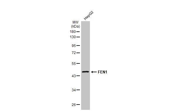
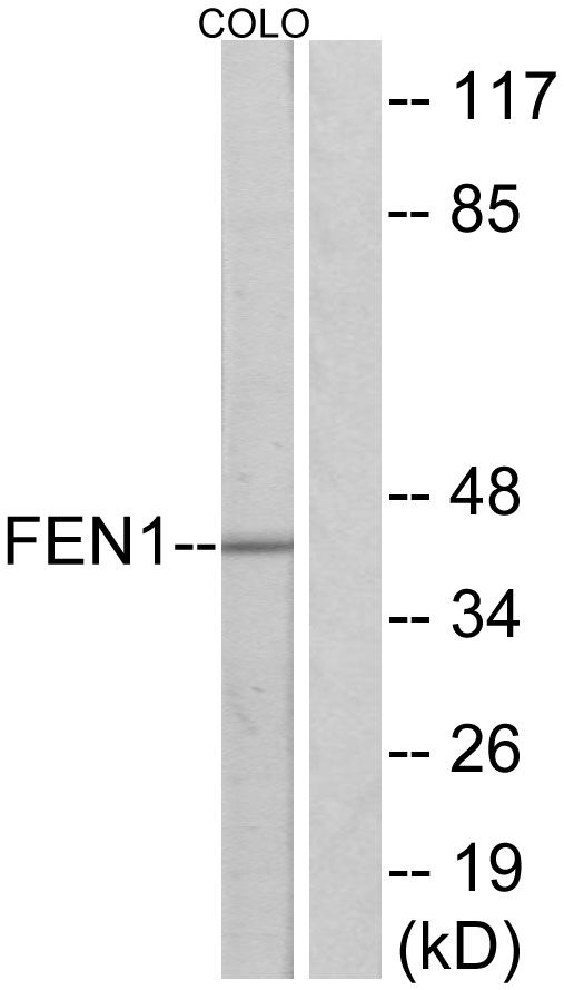
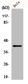
Whole cell extract (30 μg) was separated by 10% SDS-PAGE, and the membrane was blotted with FEN1 antibody (GTX101777) diluted at 1:1000. The HRP-conjugated anti-rabbit IgG antibody (GTX213110-01) was used to detect the primary antibody, and the signal was developed with Trident ECL plus-Enhanced.
![FEN1 antibody immunoprecipitates FEN1 protein in IP experiments. IP samples: Jurkat whole cell extract A. 30 μg Jurkat whole cell extract B. Control with 4 μg of preimmune Rabbit IgG C. Immunoprecipitation of FEN1 protein by 4 μg FEN1 antibody (GTX101777) 10 % SDS-PAGE The immunoprecipitated FEN1 protein was detected by FEN1 antibody (GTX101777) diluted at 1:500. [EasyBlot anti-rabbit IgG (GTX221666-01) was used as a secondary reagent]](https://www.genetex.com/upload/website/prouct_img/normal/GTX101777/GTX101777_40093_IP_w_23060100_415.webp "FEN1 antibody immunoprecipitates FEN1 protein in IP experiments. IP samples: Jurkat whole cell extract A. 30 μg Jurkat whole cell extract B. Control with 4 μg of preimmune Rabbit IgG C. Immunoprecipitation of FEN1 protein by 4 μg FEN1 antibody (GTX101777) 10 % SDS-PAGE The immunoprecipitated FEN1 protein was detected by FEN1 antibody (GTX101777) diluted at 1:500. [EasyBlot anti-rabbit IgG (GTX221666-01) was used as a secondary reagent]")
 dilution: 1:1000 The HRP-conjugated anti-rabbit IgG antibody (GTX213110-01) was used to detect the primary antibody.")
 dilution: 1:1000 The HRP-conjugated anti-rabbit IgG antibody (GTX213110-01) was used to detect the primary antibody.")
![FEN1 antibody detects FEN1 protein at nucleus by immunofluorescent analysis. Sample: HeLa cells were fixed in 4% paraformaldehyde at RT for 15 min. Green: FEN1 stained by FEN1 antibody (GTX101777) diluted at 1:500. Red: alpha Tubulin, a cytoskeleton marker, stained by alpha Tubulin antibody [GT114] (GTX628802) diluted at 1:1000. Blue: Fluoroshield with DAPI (GTX30920).](https://www.genetex.com/upload/website/prouct_img/normal/GTX101777/GTX101777_44279_20220121_ICC_IF_w_23060100_777.webp "FEN1 antibody detects FEN1 protein at nucleus by immunofluorescent analysis. Sample: HeLa cells were fixed in 4% paraformaldehyde at RT for 15 min. Green: FEN1 stained by FEN1 antibody (GTX101777) diluted at 1:500. Red: alpha Tubulin, a cytoskeleton marker, stained by alpha Tubulin antibody [GT114] (GTX628802) diluted at 1:1000. Blue: Fluoroshield with DAPI (GTX30920).")
 antibody at 1:200 dilution.")
 diluted at 1:500.
Antigen Retrieval: Trilogy? (EDTA based, pH 8.0) buffer, 15min")
Whole cell extract (30 μg) was separated by 10% SDS-PAGE, and the membrane was blotted with FEN1 antibody (GTX101777) diluted at 1:1000. The HRP-conjugated anti-rabbit IgG antibody (GTX213110-01) was used to detect the primary antibody, and the signal was developed with Trident ECL plus-Enhanced.
FEN1 antibody
GTX101777
ApplicationsImmunoFluorescence, ImmunoPrecipitation, Western Blot, ImmunoCytoChemistry, ImmunoHistoChemistry, ImmunoHistoChemistry Paraffin, Other Application
Product group Antibodies
ReactivityHuman, Mouse, Rat
TargetFEN1
Overview
- SupplierGeneTex
- Product NameFEN1 antibody
- Delivery Days Customer9
- Application Supplier NoteWB: 1:500-1:3000. ICC/IF: 1:100-1:1000. IHC-P: 1:100-1:1000. IP: 1:100-1:500. *Optimal dilutions/concentrations should be determined by the researcher.Not tested in other applications.
- ApplicationsImmunoFluorescence, ImmunoPrecipitation, Western Blot, ImmunoCytoChemistry, ImmunoHistoChemistry, ImmunoHistoChemistry Paraffin, Other Application
- CertificationResearch Use Only
- ClonalityPolyclonal
- Concentration0.2 mg/ml
- ConjugateUnconjugated
- Gene ID2237
- Target nameFEN1
- Target descriptionflap structure-specific endonuclease 1
- Target synonymsFEN-1, MF1, RAD2, flap endonuclease 1, DNase IV, maturation factor-1
- HostRabbit
- IsotypeIgG
- Protein IDP39748
- Protein NameFlap endonuclease 1
- Scientific DescriptionThe protein encoded by this gene removes 5 overhanging flaps in DNA repair and processes the 5 ends of Okazaki fragments in lagging strand DNA synthesis. Direct physical interaction between this protein and AP endonuclease 1 during long-patch base excision repair provides coordinated loading of the proteins onto the substrate, thus passing the substrate from one enzyme to another. The protein is a member of the XPG/RAD2 endonuclease family and is one of ten proteins essential for cell-free DNA replication. DNA secondary structure can inhibit flap processing at certain trinucleotide repeats in a length-dependent manner by concealing the 5 end of the flap that is necessary for both binding and cleavage by the protein encoded by this gene. Therefore, secondary structure can deter the protective function of this protein, leading to site-specific trinucleotide expansions. [provided by RefSeq]
- ReactivityHuman, Mouse, Rat
- Storage Instruction-20°C or -80°C,2°C to 8°C
- UNSPSC41116161
Datasheet
Related products
Product group Antibodies
Anti-FEN1 AntibodyA95369
ApplicationsImmunoFluorescence, Western Blot, ELISA, ImmunoHistoChemistry
ReactivityHuman, Mouse, Rat
- SizePrice
Product group Antibodies

Anti-FEN1 [SAIC-21C-4]Ab00311-1.1
ApplicationsMass Spectrometry
ReactivityHuman
TargetFEN1
- SizePrice
Product group Antibodies
Anti-FEN1 Antibody144-01175
ApplicationsImmunoFluorescence, ImmunoPrecipitation, Western Blot, ImmunoHistoChemistry
ReactivityHuman, Mouse
TargetFEN1
- SizePrice

Product group Antibodies
Anti-FEN1 Antibody Picoband(r)A01484-1-CARRIER-FREE
ApplicationsFlow Cytometry, ImmunoFluorescence, Western Blot, ELISA, ImmunoCytoChemistry, ImmunoHistoChemistry
ReactivityHuman, Mouse, Rat
TargetFEN1
- SizePrice
Product group Antibodies
FEN1 Recombinant Antibody, AbBy Fluor-594 ConjugatedBSM-61550R-BF594
ApplicationsImmunoFluorescence, Western Blot
ReactivityHuman, Mouse, Rat
TargetFEN1
- SizePrice
Product group Antibodies
FEN1 AntibodyCSB-PA002510
ApplicationsImmunoFluorescence, Western Blot, ELISA, ImmunoHistoChemistry
ReactivityHuman, Mouse, Rat
TargetFEN1
- SizePrice
Product group Antibodies
Fen1 Polyclonal AntibodyCAC11682
ApplicationsWestern Blot, ELISA
ReactivityPlant
- SizePrice
Product group Antibodies

FEN1 AntibodyLS-C331322
ApplicationsImmunoFluorescence, ImmunoPrecipitation, Western Blot, ImmunoHistoChemistry
ReactivityHuman, Mouse
TargetFEN1
- SizePrice

Product group Antibodies
Anti-FEN1 AntibodyHPA006748
ApplicationsWestern Blot, ImmunoHistoChemistry
ReactivityHuman, Mouse, Rat
TargetFEN1
- SizePrice
![ICC/IF analysis of 4% PFA-fixed HeLa cells using GTX66841 FEN1 antibody [7H8]. Dilution : 1:400](https://www.genetex.com/upload/website/prouct_img/normal/GTX66841/GTX66841_20190305_ICCIF_w_23061221_171.webp)
Product group Antibodies
FEN1 antibody [7H8]GTX66841
ApplicationsImmunoFluorescence, Western Blot, ImmunoCytoChemistry
ReactivityHamster, Human, Monkey, Mouse, Rat
TargetFEN1
- SizePrice