

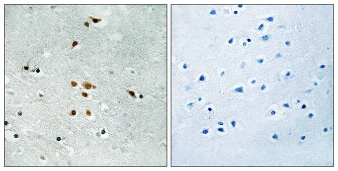
IHC-P analysis of human brain tissue using GTX55330 FER (phospho Tyr402) antibody. Left : Primary antibody Right : Primary antibody pre-incubated with the antigen specific peptide
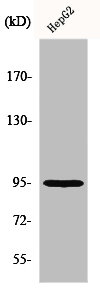
 and COS-7 cells (lane 3) using GTX55330 FER (phospho Tyr402) antibody. Left : Primary antibody pre-incubated with the antigen specific peptide Right : Primary antibody")
IHC-P analysis of human brain tissue using GTX55330 FER (phospho Tyr402) antibody. Left : Primary antibody Right : Primary antibody pre-incubated with the antigen specific peptide
FER (phospho Tyr402) antibody
GTX55330
ApplicationsWestern Blot, ImmunoHistoChemistry, ImmunoHistoChemistry Paraffin
Product group Antibodies
ReactivityHuman, Monkey
TargetFER
Overview
- SupplierGeneTex
- Product NameFER (phospho Tyr402) antibody
- Delivery Days Customer9
- Application Supplier NoteWB: 1:500-1:1000. IHC-P: 1:50-1:100. *Optimal dilutions/concentrations should be determined by the researcher.Not tested in other applications.
- ApplicationsWestern Blot, ImmunoHistoChemistry, ImmunoHistoChemistry Paraffin
- CertificationResearch Use Only
- ClonalityPolyclonal
- Concentration1 mg/ml
- ConjugateUnconjugated
- Gene ID2241
- Target nameFER
- Target descriptionFER tyrosine kinase
- Target synonymsPPP1R74, TYK3, p94-Fer, tyrosine-protein kinase Fer, feline encephalitis virus-related kinase FER, fer (fps/fes related) tyrosine kinase, fujinami poultry sarcoma/Feline sarcoma-related protein Fer, phosphoprotein NCP94, protein phosphatase 1, regulatory subunit 74, proto-oncogene c-Fer, tyrosine kinase 3
- HostRabbit
- IsotypeIgG
- Protein IDP16591
- Protein NameTyrosine-protein kinase Fer
- Scientific DescriptionThe protein encoded by this gene is a member of the FPS/FES family of non-transmembrane receptor tyrosine kinases. It regulates cell-cell adhesion and mediates signaling from the cell surface to the cytoskeleton via growth factor receptors. Alternative splicing results in multiple transcript variants. A related pseudogene has been identified on chromosome X. [provided by RefSeq, Apr 2015]
- ReactivityHuman, Monkey
- Storage Instruction-20°C or -80°C,2°C to 8°C
- UNSPSC41116161
Datasheet
Related products
Product group Antibodies
FER AntibodyCSB-PA002512
ApplicationsImmunoFluorescence, Western Blot, ELISA, ImmunoHistoChemistry
ReactivityHuman, Mouse, Rat
TargetFER
- SizePrice

Product group Antibodies
Anti-FER Antibody Picoband(r)A01630-3-CARRIER-FREE
ApplicationsWestern Blot, ELISA
ReactivityHuman, Mouse, Rat
TargetFER
- SizePrice
Product group Antibodies
Anti-FER AntibodyA98520
ApplicationsWestern Blot, ELISA
ReactivityHuman, Mouse, Rat
- SizePrice
Product group Antibodies

FER AntibodyLS-C830074
ApplicationsELISA, ImmunoHistoChemistry
ReactivityHuman, Mouse, Rat
TargetFER
- SizePrice

Product group Antibodies
Anti-FER AntibodyHPA007641
ApplicationsImmunoCytoChemistry, ImmunoHistoChemistry
ReactivityHuman
TargetFER
- SizePrice
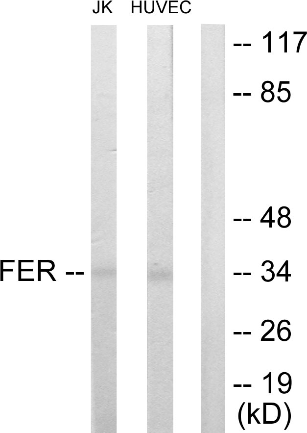
![WB analysis of NIH3T3 (1), A549 (2) and SK-MEL-5 (3) cell lysate using GTX83329 FER antibody [5D2C4].](https://www.genetex.com/upload/website/prouct_img/normal/GTX83329/GTX83329_20170912_WB_w_23061322_224.webp)
Product group Antibodies
FER antibody [5D2C4]GTX83329
ApplicationsImmunoFluorescence, Western Blot, ELISA, ImmunoCytoChemistry, ImmunoHistoChemistry, ImmunoHistoChemistry Paraffin
ReactivityHuman, Mouse
TargetFER
- SizePrice
Product group Antibodies
FER antibodyGTX12150
ApplicationsWestern Blot, ImmunoHistoChemistry, ImmunoHistoChemistry Paraffin
ReactivityHuman, Rat
TargetFER
- SizePrice
Product group Antibodies
Anti-FER Antibody144-09687
ApplicationsWestern Blot
ReactivityHuman, Mouse, Rat
TargetFER
- SizePrice
Product group Antibodies
FER Polyclonal AntibodyBS-6914R
ApplicationsImmunoFluorescence, Western Blot, ELISA, ImmunoCytoChemistry, ImmunoHistoChemistry, ImmunoHistoChemistry Frozen, ImmunoHistoChemistry Paraffin
ReactivityBovine, Canine, Equine, Human, Mouse, Porcine, Rabbit, Rat, Sheep
TargetFER
- SizePrice