

The image on the left is immunohistochemistry of paraffin-embedded Human thyroid cancer tissue using CSB-PA049381(FHIT Antibody) at dilution 1/25, on the right is treated with fusion protein. (Original magnification: x200)
 at dilution 1/350, Secondary antibody: Goat anti rabbit IgG at 1/8000 dilution, Exposure time: 30 seconds")
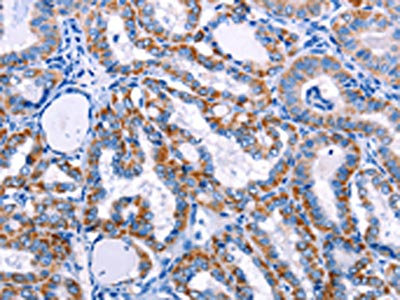
The image on the left is immunohistochemistry of paraffin-embedded Human thyroid cancer tissue using CSB-PA049381(FHIT Antibody) at dilution 1/25, on the right is treated with fusion protein. (Original magnification: x200)
FHIT Antibody
CSB-PA049381
ApplicationsWestern Blot, ELISA, ImmunoHistoChemistry
Product group Antibodies
ReactivityHuman, Mouse, Rat
TargetFHIT
Overview
- SupplierCusabio
- Product NameFHIT Antibody
- Delivery Days Customer20
- ApplicationsWestern Blot, ELISA, ImmunoHistoChemistry
- CertificationResearch Use Only
- ClonalityPolyclonal
- ConjugateUnconjugated
- Gene ID2272
- Target nameFHIT
- Target descriptionfragile histidine triad diadenosine triphosphatase
- Target synonymsAP3Aase, FRA3B, bis(5'-adenosyl)-triphosphatase, AP3A hydrolase, adenosine 5'-monophosphoramidase FHIT, adenylylsulfatase, adenylylsulfate-ammonia adenylyltransferase, diadenosine 5',5'''-P1,P3-triphosphate hydrolase, dinucleosidetriphosphatase
- HostRabbit
- IsotypeIgG
- Protein IDP49789
- Protein NameBis(5'-adenosyl)-triphosphatase
- Scientific DescriptionThis gene, a member of the histidine triad gene family, encodes a diadenosine 5,5-P1,P3-triphosphate hydrolase involved in purine metabolism. The gene encompasses the common fragile site FRA3B on chromosome 3, where carcinogen-induced damage can lead to translocations and aberrant transcripts of this gene. In fact, aberrant transcripts from this gene have been found in about half of all esophageal, stomach, and colon carcinomas. Alternatively spliced transcript variants have been found for this gene.
- ReactivityHuman, Mouse, Rat
- Storage Instruction-20°C or -80°C
- UNSPSC41116161
Related products
Product group Antibodies
Anti-FHIT AntibodyA101270
ApplicationsELISA, ImmunoHistoChemistry
ReactivityHuman
- SizePrice

Product group Antibodies
Anti-FHIT AntibodyHPA018909
ApplicationsWestern Blot, ImmunoHistoChemistry
ReactivityHuman
TargetFHIT
- SizePrice
Product group Antibodies

FHIT AntibodyLS-C401681
ApplicationsWestern Blot, ELISA, ImmunoHistoChemistry
ReactivityHuman, Mouse, Rat
TargetFHIT
- SizePrice

Product group Antibodies
Anti-FHIT Antibody Picoband(r)PB9181-CARRIER-FREE
ApplicationsFlow Cytometry, ImmunoFluorescence, Western Blot, ImmunoCytoChemistry, ImmunoHistoChemistry
ReactivityHuman, Rat
TargetFHIT
- SizePrice
Product group Antibodies
FHIT Polyclonal AntibodyBS-1769R
ApplicationsImmunoFluorescence, Western Blot, ELISA, ImmunoCytoChemistry, ImmunoHistoChemistry, ImmunoHistoChemistry Frozen, ImmunoHistoChemistry Paraffin
ReactivityBovine, Canine, Chicken, Equine, Human, Mouse, Porcine, Rabbit, Rat
TargetFHIT
- SizePrice
![Various whole cell extracts (30 μg) were separated by 15% SDS-PAGE, and the membrane was blotted with FHIT antibody [GT1240] (GTX02837) diluted at 1:1000. The HRP-conjugated anti-rabbit IgG antibody (GTX213110-01) was used to detect the primary antibody.](https://www.genetex.com/upload/website/prouct_img/normal/GTX02837/GTX02837_4000001401_20210122_WB_w_23053123_527.webp)
Product group Antibodies
FHIT antibody [GT1240]GTX02837
ApplicationsWestern Blot
ReactivityHuman, Rat
TargetFHIT
- SizePrice
Product group Antibodies
Anti-FHIT Antibody144-01196
ApplicationsWestern Blot, ImmunoHistoChemistry
ReactivityHuman, Mouse, Rat
TargetFHIT
- SizePrice