

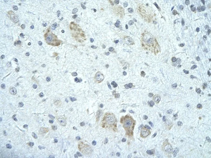
IHC-P analysis of human brain tissue using GTX47245 FIP1L1 antibody at 4.0-8.0μg/ml.

IHC-P analysis of human brain tissue using GTX47245 FIP1L1 antibody at 4.0-8.0μg/ml.
FIP1L1 antibody, C-term
GTX47245
ApplicationsWestern Blot, ImmunoHistoChemistry, ImmunoHistoChemistry Paraffin
Product group Antibodies
ReactivityHuman
TargetFIP1L1
Overview
- SupplierGeneTex
- Product NameFIP1L1 antibody, C-term
- Delivery Days Customer9
- Application Supplier NoteWB: 0.2-2.5 ug/ml. IHC-P: 2-10 ug/ml. *Optimal dilutions/concentrations should be determined by the researcher.Not tested in other applications.
- ApplicationsWestern Blot, ImmunoHistoChemistry, ImmunoHistoChemistry Paraffin
- CertificationResearch Use Only
- ClonalityPolyclonal
- Concentration0.5-1 mg/ml
- ConjugateUnconjugated
- Gene ID81608
- Target nameFIP1L1
- Target descriptionfactor interacting with PAPOLA and CPSF1
- Target synonymsFIP1, Rhe, hFip1, pre-mRNA 3'-end-processing factor FIP1, FIP1 like 1, FIP1-like 1 protein, FIP1L1 cleavage and polyadenylation specific factor subunit, factor interacting with PAP, rearranged in hypereosinophilia
- HostRabbit
- IsotypeIgG
- Protein IDQ6UN15
- Protein NamePre-mRNA 3'-end-processing factor FIP1
- Scientific DescriptionThis gene encodes a subunit of the CPSF (cleavage and polyadenylation specificity factor) complex that polyadenylates the 3 end of mRNA precursors. This gene, the homolog of yeast Fip1 (factor interacting with PAP), binds to U-rich sequences of pre-mRNA and stimulates poly(A) polymerase activity. Its N-terminus contains a PAP-binding site and its C-terminus an RNA-binding domain. An interstitial chromosomal deletion on 4q12 creates an in-frame fusion of human genes FIP1L1 and PDGFRA (platelet-derived growth factor receptor, alpha). The FIP1L1-PDGFRA fusion gene encodes a constitutively activated tyrosine kinase that joins the first 233 amino acids of FIP1L1 to the last 523 amino acids of PDGFRA. This gene fusion and chromosomal deletion is the cause of some forms of idiopathic hypereosinophilic syndrome (HES). This syndrome, recently reclassified as chronic eosinophilic leukemia (CEL), is responsive to treatment with tyrosine kinase inhibitors. Alternative splicing results in multiple transcript variants encoding distinct isoforms. [provided by RefSeq, Oct 2008]
- ReactivityHuman
- Storage Instruction-20°C or -80°C,2°C to 8°C
- UNSPSC41116161
Datasheet
Related products
Product group Antibodies
Anti-FIP1L1 AntibodyA31903
ApplicationsImmunoFluorescence, Western Blot, ImmunoHistoChemistry
ReactivityHuman, Mouse, Rat
- SizePrice

Product group Antibodies
Anti-FIP1L1 Antibody Picoband(r)A02452-1-CARRIER-FREE
ApplicationsFlow Cytometry, ImmunoFluorescence, Western Blot, ELISA, ImmunoCytoChemistry, ImmunoHistoChemistry
ReactivityHuman, Mouse, Rat
TargetFIP1L1
- SizePrice
Product group Antibodies
Anti-FIP1L1 Antibody144-07138
ApplicationsImmunoFluorescence, Western Blot, ImmunoHistoChemistry
ReactivityHuman, Mouse, Rat
TargetFIP1L1
- SizePrice
Product group Antibodies
FIP1L1 Polyclonal AntibodyBS-13173R
ApplicationsImmunoFluorescence, Western Blot, ELISA, ImmunoCytoChemistry, ImmunoHistoChemistry, ImmunoHistoChemistry Frozen, ImmunoHistoChemistry Paraffin
ReactivityBovine, Canine, Chicken, Equine, Human, Mouse, Porcine, Rabbit, Rat, Sheep
TargetFIP1L1
- SizePrice
Product group Antibodies

HFip1 / FIP1L1 AntibodyLS-C346244
ApplicationsImmunoFluorescence, Western Blot, ImmunoHistoChemistry
ReactivityHuman, Mouse, Rat
TargetFIP1L1
- SizePrice
Product group Antibodies
FIP1L1 antibodyGTX55623
ApplicationsImmunoFluorescence, Western Blot, ImmunoCytoChemistry, ImmunoHistoChemistry, ImmunoHistoChemistry Paraffin
ReactivityHuman, Mouse, Rat
TargetFIP1L1
- SizePrice

Product group Antibodies
Anti-FIP1L1 AntibodyHPA037475
ApplicationsWestern Blot, ImmunoCytoChemistry, ImmunoHistoChemistry
ReactivityHuman
TargetFIP1L1
- SizePrice
Product group Antibodies
Anti-FIP1L1 AntibodyCAB7138
ApplicationsImmunoFluorescence, Western Blot, ELISA, ImmunoCytoChemistry, ImmunoHistoChemistry, ImmunoHistoChemistry Paraffin
ReactivityHuman
TargetFIP1L1
- SizePrice