

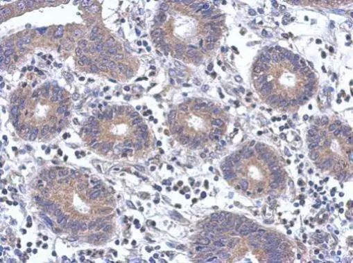
Immunohistochemical analysis of paraffin-embedded human colon carcinoma, using FYN(GTX109428) antibody at 1:500 dilution.
Antigen Retrieval: Trilogy? (EDTA based, pH 8.0) buffer, 15min
 A: Jurkat 10% SDS PAGE GTX109428 diluted at 1:1000")
![FYN antibody [N1C2] detects FYN protein by western blot analysis. Mouse and rat tissue extracts (50 μg) were separated by 10% SDS-PAGE, and the membrane was blotted with FYN antibody [N1C2] (GTX109428) diluted at 1:1000.](https://www.genetex.com/upload/website/prouct_img/normal/GTX109428/GTX109428_40394_20151022_WB_Merged_w_23060500_979.webp "FYN antibody [N1C2] detects FYN protein by western blot analysis. Mouse and rat tissue extracts (50 μg) were separated by 10% SDS-PAGE, and the membrane was blotted with FYN antibody [N1C2] (GTX109428) diluted at 1:1000.")
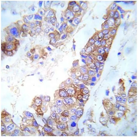
Immunohistochemical analysis of paraffin-embedded human colon carcinoma, using FYN(GTX109428) antibody at 1:500 dilution.
Antigen Retrieval: Trilogy? (EDTA based, pH 8.0) buffer, 15min
FYN antibody [N1C2]
GTX109428
ApplicationsWestern Blot, ImmunoHistoChemistry, ImmunoHistoChemistry Paraffin
Product group Antibodies
ReactivityHuman, Mouse, Rat
TargetFYN
Overview
- SupplierGeneTex
- Product NameFYN antibody [N1C2]
- Delivery Days Customer9
- Application Supplier NoteWB: 1:500-1:3000. IHC-P: 1:100-1:1000. *Optimal dilutions/concentrations should be determined by the researcher.Not tested in other applications.
- ApplicationsWestern Blot, ImmunoHistoChemistry, ImmunoHistoChemistry Paraffin
- CertificationResearch Use Only
- ClonalityPolyclonal
- Concentration1.01 mg/ml
- ConjugateUnconjugated
- Gene ID2534
- Target nameFYN
- Target descriptionFYN proto-oncogene, Src family tyrosine kinase
- Target synonymsSLK, SYN, p59-FYN, tyrosine-protein kinase Fyn, FYN oncogene related to SRC, FGR, YES, OKT3-induced calcium influx regulator, c-syn protooncogene, proto-oncogene Syn, proto-oncogene c-Fyn, src-like kinase, src/yes-related novel, tyrosine kinase p59fyn(T)
- HostRabbit
- IsotypeIgG
- Protein IDP06241
- Protein NameTyrosine-protein kinase Fyn
- Scientific DescriptionThis gene is a member of the protein-tyrosine kinase oncogene family. It encodes a membrane-associated tyrosine kinase that has been implicated in the control of cell growth. The protein associates with the p85 subunit of phosphatidylinositol 3-kinase and interacts with the fyn-binding protein. Alternatively spliced transcript variants encoding distinct isoforms exist. [provided by RefSeq]
- ReactivityHuman, Mouse, Rat
- Storage Instruction-20°C or -80°C,2°C to 8°C
- UNSPSC12352203
References
- Lee JH, Ostalecki C, Zhao Z, et al. HIV Activates the Tyrosine Kinase Hck to Secrete ADAM Protease-Containing Extracellular Vesicles. EBioMedicine. 2018,28:151-161. doi: 10.1016/j.ebiom.2018.01.004Read this paper
Datasheet
Related products
Product group Antibodies
ApplicationsImmunoPrecipitation, Western Blot
ReactivityHuman
TargetFYN
- SizePrice

Product group Antibodies
ApplicationsImmunoFluorescence, Western Blot, ImmunoCytoChemistry
ReactivityHuman, Mouse, Rat
TargetFYN
- SizePrice
![Whole zebrafish extract (30 μg) was separated by 7.5% SDS-PAGE, and the membrane was blotted with FYN antibody [N1N3] (GTX101189) diluted at 1:1000.](https://www.genetex.com/upload/website/prouct_img/normal/GTX101189/GTX101189_39617_20170209_WB_Z_22111422_732.webp)
Product group Antibodies
References
FYN antibody [N1N3]GTX101189
ApplicationsImmunoFluorescence, Western Blot, ImmunoCytoChemistry, ImmunoHistoChemistry
ReactivityHuman, Mouse, Zebra Fish
TargetFYN
- SizePrice
![FACS analysis of HeLa cells using GTX60460 FYN antibody [2A10]. Green : FYN Purple : negative control](https://www.genetex.com/upload/website/prouct_img/normal/GTX60460/GTX60460_20170912_FACS_w_23061123_958.webp)
Product group Antibodies
FYN antibody [2A10]GTX60460
ApplicationsFlow Cytometry, ImmunoFluorescence, Western Blot, ELISA, ImmunoCytoChemistry, ImmunoHistoChemistry, ImmunoHistoChemistry Paraffin
ReactivityHuman
TargetFYN
- SizePrice
![ELISA analysis of antigen using GTX60461 FYN antibody [2H8].
Red : Control antigen 100ng
Purple : Antigen 10ng
Green : Antigen 50ng
Blue : Antigen 100ng](https://www.genetex.com/upload/website/prouct_img/normal/GTX60461/GTX60461_20170912_ELISA_w_23061123_465.webp)
Product group Antibodies
FYN antibody [2H8]GTX60461
ApplicationsWestern Blot, ELISA
ReactivityHuman, Mouse
TargetFYN
- SizePrice
Product group Antibodies
FYN (phospho Tyr530) antibodyGTX79071
ApplicationsImmunoHistoChemistry, ImmunoHistoChemistry Paraffin
ReactivityHuman
TargetFYN
- SizePrice
Product group Antibodies
FYN (phospho Tyr530) antibodyGTX86757
ApplicationsWestern Blot, ImmunoHistoChemistry, ImmunoHistoChemistry Paraffin
ReactivityHuman, Mouse
TargetFYN
- SizePrice
Product group Antibodies
Fyn Polyclonal AntibodyCAC08453
ApplicationsImmunoFluorescence, ELISA, ImmunoHistoChemistry
TargetFYN
- SizePrice
Product group Antibodies
FYN Recombinant AntibodyBSM-61140R
ApplicationsImmunoFluorescence, Western Blot, ImmunoCytoChemistry
TargetFYN
- SizePrice