

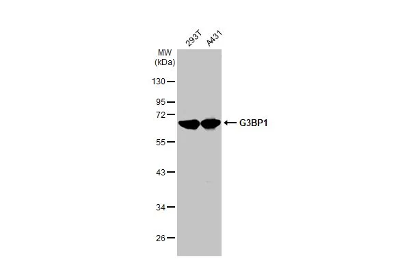
Various whole cell extracts (30 μg) were separated by 10% SDS-PAGE, and the membrane was blotted with G3BP1 antibody (GTX112191) diluted at 1:5000. The HRP-conjugated anti-rabbit IgG antibody (GTX213110-01) was used to detect the primary antibody.
 diluted at 1:2000. Blue: Fluoroshield with DAPI (GTX30920). Scale bar= 10 μm.")
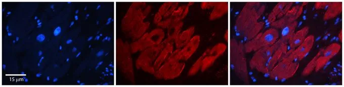
 diluted at 1:250. Antigen Retrieval: Tris-EDTA buffer, pH 9.0, 15 min")
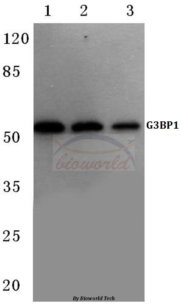
Various whole cell extracts (30 μg) were separated by 10% SDS-PAGE, and the membrane was blotted with G3BP1 antibody (GTX112191) diluted at 1:5000. The HRP-conjugated anti-rabbit IgG antibody (GTX213110-01) was used to detect the primary antibody.
G3BP1 antibody
GTX112191
ApplicationsImmunoFluorescence, Western Blot, ImmunoCytoChemistry, ImmunoHistoChemistry, ImmunoHistoChemistry Paraffin
Product group Antibodies
ReactivityHuman, Mouse
TargetG3BP1
Overview
- SupplierGeneTex
- Product NameG3BP1 antibody
- Delivery Days Customer9
- Application Supplier NoteWB: 1:1000-1:10000. ICC/IF: 1:100-1:1000. IHC-P: 1:100-1:1000. *Optimal dilutions/concentrations should be determined by the researcher.Not tested in other applications.
- ApplicationsImmunoFluorescence, Western Blot, ImmunoCytoChemistry, ImmunoHistoChemistry, ImmunoHistoChemistry Paraffin
- CertificationResearch Use Only
- ClonalityPolyclonal
- Concentration0.35 mg/ml
- ConjugateUnconjugated
- Gene ID10146
- Target nameG3BP1
- Target descriptionG3BP stress granule assembly factor 1
- Target synonymsG3BP, HDH-VIII, ras GTPase-activating protein-binding protein 1, ATP-dependent DNA helicase VIII, DNA helicase VIII, G3BP-1, GAP SH3 domain-binding protein 1, GAP binding protein, GTPase activating protein (SH3 domain) binding protein 1, Ras-GTPase-activating protein SH3-domain-binding protein, RasGAP-associated endoribonuclease G3BP
- HostRabbit
- IsotypeIgG
- Protein IDQ13283
- Protein NameRas GTPase-activating protein-binding protein 1
- Scientific DescriptionThis gene encodes one of the DNA-unwinding enzymes which prefers partially unwound 3-tailed substrates and can also unwind partial RNA/DNA and RNA/RNA duplexes in an ATP-dependent fashion. This enzyme is a member of the heterogeneous nuclear RNA-binding proteins and is also an element of the Ras signal transduction pathway. It binds specifically to the Ras-GTPase-activating protein by associating with its SH3 domain. Several alternatively spliced transcript variants of this gene have been described, but the full-length nature of some of these variants has not been determined. [provided by RefSeq]
- ReactivityHuman, Mouse
- Storage Instruction-20°C or -80°C,2°C to 8°C
- UNSPSC41116161
Datasheet
Related products
Product group Antibodies
ApplicationsWestern Blot, ImmunoHistoChemistry
ReactivityHuman, Mouse
- SizePrice
Product group Antibodies
Anti-G3BP1 Antibody144-60815
ApplicationsWestern Blot, ImmunoHistoChemistry
ReactivityHuman, Mouse, Rat
TargetG3BP1
- SizePrice
Product group Antibodies

G3BP1 / G3BP AntibodyLS-C749818
ApplicationsWestern Blot
ReactivityHuman
TargetG3BP1
- SizePrice

Product group Antibodies
Anti-G3BP/G3BP1 Antibody Picoband(r)A02199-2-CARRIER-FREE
ApplicationsFlow Cytometry, ImmunoFluorescence, Western Blot, ImmunoCytoChemistry, ImmunoHistoChemistry
ReactivityHuman
TargetG3BP1
- SizePrice
Product group Antibodies
G3BP1 Recombinant Antibody, AbBy Fluor-555 ConjugatedBSM-62130R-BF555
ApplicationsFlow Cytometry, ImmunoFluorescence, Western Blot
ReactivityHuman, Mouse, Rat
TargetG3BP1
- SizePrice
Product group Antibodies
G3BP1 AntibodyCSB-PA002599
ApplicationsWestern Blot, ELISA, ImmunoHistoChemistry
ReactivityHuman, Monkey, Mouse
TargetG3BP1
- SizePrice
Product group Antibodies
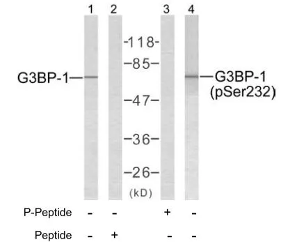
G3BP1 (phospho Ser232) antibodyGTX79070
ApplicationsWestern Blot, ImmunoHistoChemistry, ImmunoHistoChemistry Paraffin
ReactivityHuman
TargetG3BP1
- SizePrice

Product group Antibodies
Anti-G3BP1 AntibodyHPA004052
ApplicationsWestern Blot, ImmunoCytoChemistry, ImmunoHistoChemistry
ReactivityHuman, Mouse, Rat
TargetG3BP1
- SizePrice
Product group Antibodies
G3BP1 antibody, N-termGTX47440
ApplicationsImmunoPrecipitation, Western Blot, ImmunoHistoChemistry, ImmunoHistoChemistry Paraffin
ReactivityHuman, Mouse, Rat
TargetG3BP1
- SizePrice