

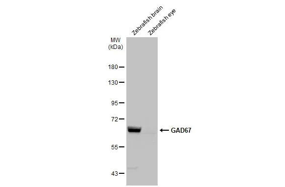
Various tissue extracts (30 μg) were separated by 7.5% SDS-PAGE, and the membrane was blotted with GAD67 antibody (GTX101881) diluted at 1:500. The HRP-conjugated anti-rabbit IgG antibody (GTX213110-01) was used to detect the primary antibody.
 dilution: 1:200. Image provided with permission courtesy of Dr. T. Schilling at UC, Irvine.")
 dilution: 1:200.")
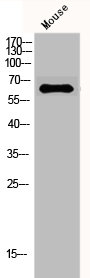
 were separated by 7.5% SDS-PAGE, and the membrane was blotted with GAD67 antibody (GTX101881) diluted at 1:1000. The HRP-conjugated anti-rabbit IgG antibody (GTX213110-01) was used to detect the primary antibody.")
 was separated by 7.5% SDS-PAGE, and the membrane was blotted with GAD67 antibody (GTX101881) diluted at 1:1000. The HRP-conjugated anti-rabbit IgG antibody (GTX213110-01) was used to detect the primary antibody.")
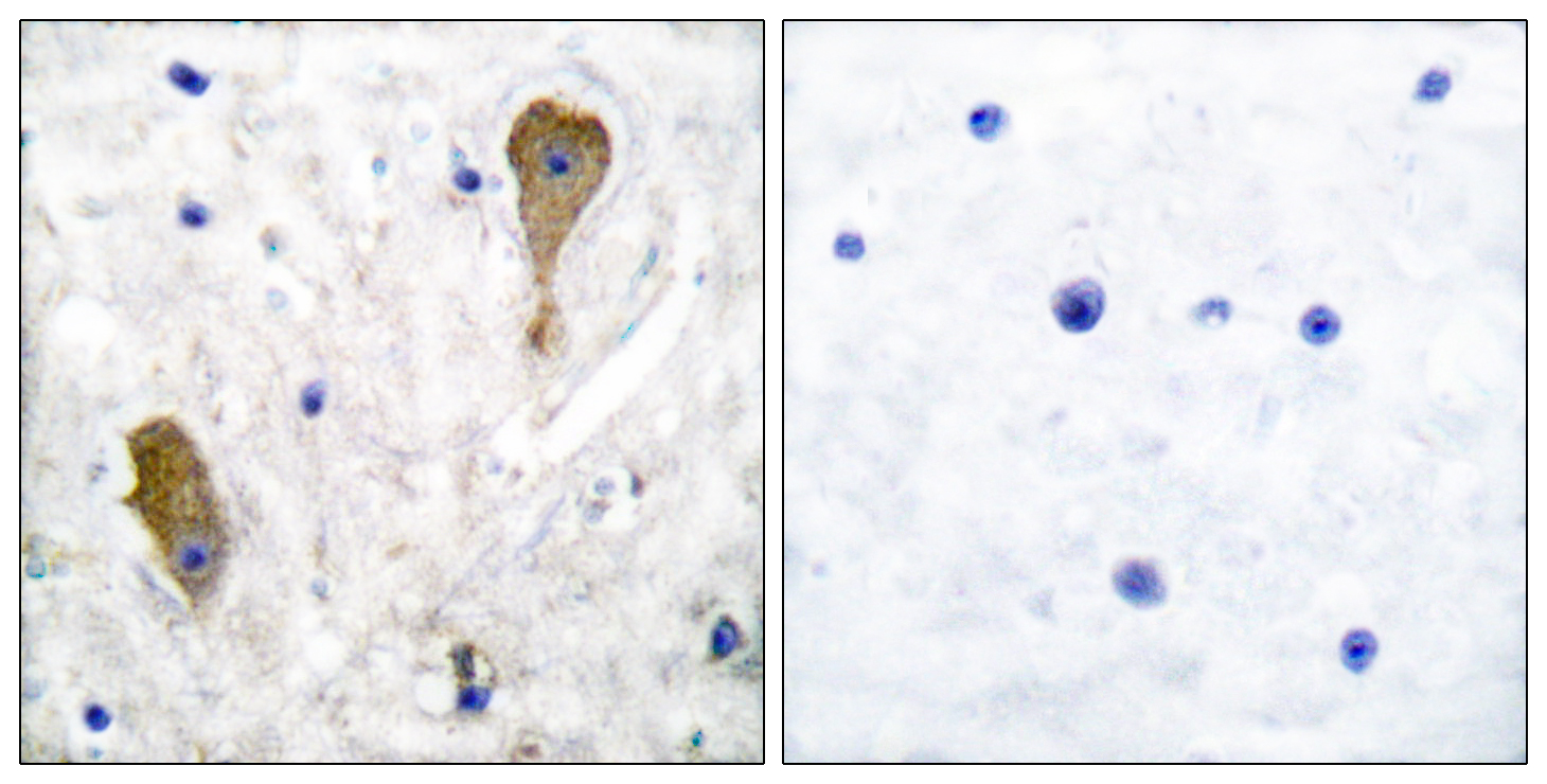
 diluted at 1:500. Antigen Retrieval: Citrate buffer, pH 6.0, 15 min")
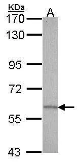
 were separated by 7.5% SDS-PAGE, and the membrane was blotted with GAD67 antibody (GTX101881) diluted at 1:500. The HRP-conjugated anti-rabbit IgG antibody (GTX213110-01) was used to detect the primary antibody.")
 diluted at 1:500. Antigen Retrieval: Citrate buffer, pH 6.0, 15 min")
 diluted at 1:500. Blue: Fluoroshield with DAPI (GTX30920).")
![GAD67 antibody detects GAD67 protein by immunofluorescent analysis. Sample: DIV10 rat E18 primary cortical neuron cells were fixed in 4% paraformaldehyde at RT for 15 min. Green: GAD67 stained by GAD67 antibody (GTX101881) diluted at 1:500. Red: Tau, stained by Tau antibody [GT287] (GTX634809) diluted at 1:500. Blue: Fluoroshield with DAPI (GTX30920).](https://www.genetex.com/upload/website/prouct_img/normal/GTX101881/GTX101881_43789_20200820_ICC_IF_R_w_23060100_538.webp "GAD67 antibody detects GAD67 protein by immunofluorescent analysis. Sample: DIV10 rat E18 primary cortical neuron cells were fixed in 4% paraformaldehyde at RT for 15 min. Green: GAD67 stained by GAD67 antibody (GTX101881) diluted at 1:500. Red: Tau, stained by Tau antibody [GT287] (GTX634809) diluted at 1:500. Blue: Fluoroshield with DAPI (GTX30920).")
Various tissue extracts (30 μg) were separated by 7.5% SDS-PAGE, and the membrane was blotted with GAD67 antibody (GTX101881) diluted at 1:500. The HRP-conjugated anti-rabbit IgG antibody (GTX213110-01) was used to detect the primary antibody.
GAD67 antibody
GTX101881
ApplicationsImmunoFluorescence, Western Blot, ImmunoCytoChemistry, ImmunoHistoChemistry, ImmunoHistoChemistry Frozen, ImmunoHistoChemistry Paraffin
Product group Antibodies
ReactivityHuman, Monkey, Mouse, Rat, Zebra Fish
TargetGAD1
Overview
- SupplierGeneTex
- Product NameGAD67 antibody
- Delivery Days Customer9
- Application Supplier NoteWB: 1:500-1:3000. ICC/IF: 1:100-1:1000. IHC-P: 1:100-1:1000. IHC-Fr: 1:100-1:1000. *Optimal dilutions/concentrations should be determined by the researcher.Not tested in other applications.
- ApplicationsImmunoFluorescence, Western Blot, ImmunoCytoChemistry, ImmunoHistoChemistry, ImmunoHistoChemistry Frozen, ImmunoHistoChemistry Paraffin
- CertificationResearch Use Only
- ClonalityPolyclonal
- Concentration0.22 mg/ml
- ConjugateUnconjugated
- Gene ID2571
- Target nameGAD1
- Target descriptionglutamate decarboxylase 1
- Target synonymsCPSQ1, DEE89, GAD, SCP, glutamate decarboxylase 1, 67 kDa glutamic acid decarboxylase, GAD-67, glutamate decarboxylase 1 (brain, 67kDa)
- HostRabbit
- IsotypeIgG
- Protein IDQ99259
- Protein NameGlutamate decarboxylase 1
- Scientific DescriptionThis gene encodes one of several forms of glutamic acid decarboxylase, identified as a major autoantigen in insulin-dependent diabetes. The enzyme encoded is responsible for catalyzing the production of gamma-aminobutyric acid from L-glutamic acid. A pathogenic role for this enzyme has been identified in the human pancreas since it has been identified as an autoantigen and an autoreactive T cell target in insulin-dependent diabetes. This gene may also play a role in the stiff man syndrome. Deficiency in this enzyme has been shown to lead to pyridoxine dependency with seizures. Alternative splicing of this gene results in two products, the predominant 67-kD form and a less-frequent 25-kD form. [provided by RefSeq]
- ReactivityHuman, Monkey, Mouse, Rat, Zebra Fish
- Storage Instruction-20°C or -80°C,2°C to 8°C
- UNSPSC41116161
Datasheet
Related products
Product group Antibodies
Anti-GAD1 AntibodyA96199
ApplicationsWestern Blot, ELISA, ImmunoHistoChemistry
ReactivityHuman, Mouse, Rat
- SizePrice
Product group Antibodies
Anti-GAD1 Antibody144-02938
ApplicationsWestern Blot
ReactivityHuman, Mouse, Rat
TargetGAD1
- SizePrice

Product group Antibodies
Anti-GAD1 AntibodyAMAB91076
ApplicationsWestern Blot, ImmunoHistoChemistry
ReactivityHuman, Mouse, Rat
TargetGAD1
- SizePrice
Product group Antibodies
References
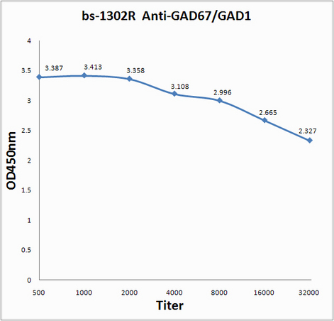
GAD67 Polyclonal AntibodyBS-1302R
ApplicationsImmunoFluorescence, Western Blot, ELISA, ImmunoCytoChemistry, ImmunoHistoChemistry, ImmunoHistoChemistry Frozen, ImmunoHistoChemistry Paraffin
ReactivityHuman, Mouse, Rat
TargetGAD1
- SizePrice
Product group Antibodies
GAD1/GAD2 AntibodyCSB-PA002622
ApplicationsImmunoFluorescence, Western Blot, ELISA, ImmunoHistoChemistry
ReactivityHuman, Mouse
TargetGAD1
- SizePrice
Product group Antibodies

ApplicationsFlow Cytometry, ImmunoFluorescence, Western Blot, ELISA
ReactivityBovine, Canine, Human, Mouse, Porcine, Rat
TargetGAD1
- SizePrice
![WB analysis of human brain tissue lysate using GTX17736 GAD67 antibody [GAD1/2391].](https://www.genetex.com/upload/website/prouct_img/normal/GTX17736/GTX17736_20200115_WB_331_w_23060620_825.webp)
Product group Antibodies
GAD67 antibody [GAD1/2391]GTX17736
ApplicationsWestern Blot, ELISA, ImmunoHistoChemistry, ImmunoHistoChemistry Paraffin, Other Application
ReactivityHuman
TargetGAD1
- SizePrice
Product group Antibodies
GAD67 antibody [N3C3]GTX101880
ApplicationsWestern Blot
ReactivityHuman
TargetGAD1
- SizePrice
![IHC-P analysis of human thyroid tissue using GTX84469 GAD67 antibody [3G9]. Antigen retrieval : Heat-induced epitope retrieval by 10mM citrate buffer, pH6.0, 100oC for 10min.](https://www.genetex.com/upload/website/prouct_img/normal/GTX84469/GTX84469_2762_IHC-P_w_23061420_461.webp)
Product group Antibodies
GAD67 antibody [3G9]GTX84469
ApplicationsImmunoFluorescence, ImmunoPrecipitation, Western Blot, ImmunoCytoChemistry, ImmunoHistoChemistry, ImmunoHistoChemistry Paraffin
ReactivityHuman, Monkey
TargetGAD1
- SizePrice