

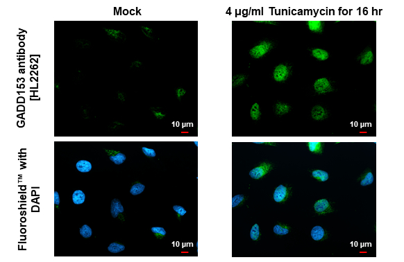
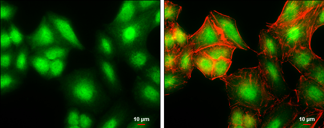
GADD153 antibody [HL2262] detects GADD153 protein at cytoplasm and nucleus by immunofluorescent analysis. Sample: Mock and treated HeLa cells were fixed in 4% paraformaldehyde at RT for 15 min. Green: GADD153 stained by GADD153 antibody [HL2262] (GTX638314) diluted at 1:500. Blue: Fluoroshield with DAPI (GTX30920).
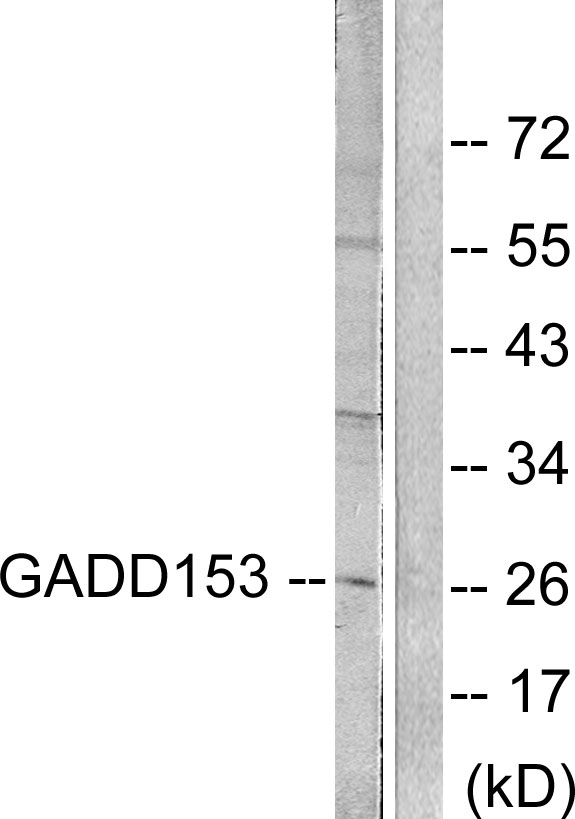
![Untreated (–) and treated (+) HepG2 whole cell extract (30 μg) were separated by 15% SDS-PAGE, and the membrane was blotted with GADD153 antibody [HL2262] (GTX638314) diluted at 1:1000. The HRP-conjugated anti-rabbit IgG antibody (GTX213110-01) was used to detect the primary antibody.](https://www.genetex.com/upload/website/prouct_img/normal/GTX638314/GTX638314_45033_20230519_WB_treatment_Tunicamycin_23053001_372.webp "Untreated (–) and treated (+) HepG2 whole cell extract (30 μg) were separated by 15% SDS-PAGE, and the membrane was blotted with GADD153 antibody [HL2262] (GTX638314) diluted at 1:1000. The HRP-conjugated anti-rabbit IgG antibody (GTX213110-01) was used to detect the primary antibody.")
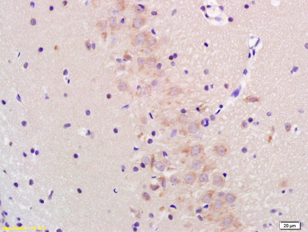
![GADD153 antibody [HL2262] detects GADD153 protein at cytoplasm and nucleus by immunohistochemical analysis. Sample: Paraffin-embedded human colon cancer. GADD153 stained by GADD153 antibody [HL2262] (GTX638314) diluted at 1:200. Antigen Retrieval: Citrate buffer, pH 6.0, 15 min](https://www.genetex.com/upload/website/prouct_img/normal/GTX638314/GTX638314_45033_20230602_IHC-P_23061320_272.webp "GADD153 antibody [HL2262] detects GADD153 protein at cytoplasm and nucleus by immunohistochemical analysis. Sample: Paraffin-embedded human colon cancer. GADD153 stained by GADD153 antibody [HL2262] (GTX638314) diluted at 1:200. Antigen Retrieval: Citrate buffer, pH 6.0, 15 min")
![GADD153 antibody [HL2262] detects GADD153 protein at cytoplasm and nucleus by immunohistochemical analysis. Sample: Paraffin-embedded human breast carcinoma. GADD153 stained by GADD153 antibody [HL2262] (GTX638314) diluted at 1:200. Antigen Retrieval: Citrate buffer, pH 6.0, 15 min](https://www.genetex.com/upload/website/prouct_img/normal/GTX638314/GTX638314_45033_20230602_IHC-P_1_23061320_101.webp "GADD153 antibody [HL2262] detects GADD153 protein at cytoplasm and nucleus by immunohistochemical analysis. Sample: Paraffin-embedded human breast carcinoma. GADD153 stained by GADD153 antibody [HL2262] (GTX638314) diluted at 1:200. Antigen Retrieval: Citrate buffer, pH 6.0, 15 min")
![Untreated (–) and treated (+) HeLa whole cell extracts (30 μg) were separated by 15% SDS-PAGE, and the membrane was blotted with GADD153 antibody [HL2262] (GTX638314) diluted at 1:4000. The HRP-conjugated anti-rabbit IgG antibody (GTX213110-01) was used to detect the primary antibody.](https://www.genetex.com/upload/website/prouct_img/normal/GTX638314/GTX638314_45125_20230804_WB_treatment_Tunicamycin_23110819_320.webp "Untreated (–) and treated (+) HeLa whole cell extracts (30 μg) were separated by 15% SDS-PAGE, and the membrane was blotted with GADD153 antibody [HL2262] (GTX638314) diluted at 1:4000. The HRP-conjugated anti-rabbit IgG antibody (GTX213110-01) was used to detect the primary antibody.")
GADD153 antibody [HL2262] detects GADD153 protein at cytoplasm and nucleus by immunofluorescent analysis. Sample: Mock and treated HeLa cells were fixed in 4% paraformaldehyde at RT for 15 min. Green: GADD153 stained by GADD153 antibody [HL2262] (GTX638314) diluted at 1:500. Blue: Fluoroshield with DAPI (GTX30920).
GADD153 antibody [HL2262]
GTX638314
ApplicationsImmunoFluorescence, Western Blot, ImmunoCytoChemistry, ImmunoHistoChemistry, ImmunoHistoChemistry Paraffin
Product group Antibodies
ReactivityHuman
TargetDDIT3
Overview
- SupplierGeneTex
- Product NameGADD153 antibody [HL2262]
- Delivery Days Customer9
- Application Supplier NoteWB: 1:1000-1:10000. *Optimal dilutions/concentrations should be determined by the researcher.Not tested in other applications.
- ApplicationsImmunoFluorescence, Western Blot, ImmunoCytoChemistry, ImmunoHistoChemistry, ImmunoHistoChemistry Paraffin
- CertificationResearch Use Only
- ClonalityMonoclonal
- Clone IDHL2262
- Concentration1 mg/ml
- ConjugateUnconjugated
- Gene ID1649
- Target nameDDIT3
- Target descriptionDNA damage inducible transcript 3
- Target synonymsAltDDIT3, C/EBPzeta, CEBPZ, CHOP, CHOP-10, CHOP10, GADD153, DNA damage-inducible transcript 3 protein, C/EBP zeta, CCAAT/enhancer-binding protein homologous protein, alternative DDIT3 protein, c/EBP-homologous protein 10, growth arrest and DNA damage-inducible protein GADD153
- HostRabbit
- IsotypeIgG
- Protein IDP35638
- Protein NameDNA damage-inducible transcript 3 protein
- Scientific DescriptionThis gene encodes a member of the CCAAT/enhancer-binding protein (C/EBP) family of transcription factors. The protein functions as a dominant-negative inhibitor by forming heterodimers with other C/EBP members, such as C/EBP and LAP (liver activator protein), and preventing their DNA binding activity. The protein is implicated in adipogenesis and erythropoiesis, is activated by endoplasmic reticulum stress, and promotes apoptosis. Fusion of this gene and FUS on chromosome 16 or EWSR1 on chromosome 22 induced by translocation generates chimeric proteins in myxoid liposarcomas or Ewing sarcoma. Multiple alternatively spliced transcript variants encoding two isoforms with different length have been identified. [provided by RefSeq, Aug 2010]
- ReactivityHuman
- Storage Instruction-20°C or -80°C,2°C to 8°C
- UNSPSC41116161
Datasheet
Related products
Product group Antibodies
DDIT3 AntibodyCSB-PA001639
ApplicationsImmunoFluorescence, Western Blot, ELISA, ImmunoHistoChemistry
ReactivityHuman, Mouse, Rat
TargetDDIT3
- SizePrice

Product group Antibodies
Anti-DDIT3 Antibody Picoband(r)A00311-2-CARRIER-FREE
ApplicationsFlow Cytometry, Western Blot, ELISA, ImmunoHistoChemistry
ReactivityHuman
TargetDDIT3
- SizePrice
Product group Antibodies
Anti-GADD153 AntibodyA94972
ApplicationsImmunoFluorescence, Western Blot, ELISA, ImmunoHistoChemistry
ReactivityHuman, Mouse, Rat
- SizePrice
Product group Antibodies

DDIT3 / CHOP Antibody (clone 1E1)LS-C765520
ApplicationsImmunoHistoChemistry, ImmunoHistoChemistry Paraffin
ReactivityHuman, Mouse, Rat
TargetDDIT3
- SizePrice

Product group Antibodies
Anti-DDIT3 AntibodyHPA058416
ApplicationsImmunoCytoChemistry
ReactivityHuman
TargetDDIT3
- SizePrice
Product group Antibodies
Ddit3 Polyclonal AntibodyCAC07500
ApplicationsImmunoFluorescence, ELISA, ImmunoHistoChemistry
TargetDDIT3
- SizePrice
Product group Antibodies
References
DDIT3 Polyclonal AntibodyBS-1361R
ApplicationsFlow Cytometry, ImmunoFluorescence, Western Blot, ELISA, ImmunoCytoChemistry, ImmunoHistoChemistry, ImmunoHistoChemistry Frozen, ImmunoHistoChemistry Paraffin
ReactivityHuman, Mouse, Rat
TargetDDIT3
- SizePrice
Product group Antibodies
GADD153 antibodyGTX109226
ApplicationsImmunoFluorescence, Western Blot, ImmunoCytoChemistry
ReactivityHuman, Mouse
TargetDDIT3
- SizePrice