

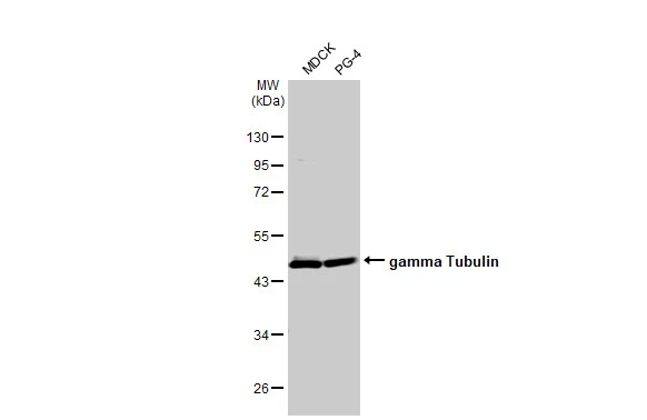
Various whole cell extracts (30 μg) were separated by 10% SDS-PAGE, and the membrane was blotted with gamma Tubulin antibody [HL1175] (GTX636480) diluted at 1:5000. The HRP-conjugated anti-rabbit IgG antibody (GTX213110-01) was used to detect the primary antibody.
![Whole cell extract (30 μg) was separated by 10% SDS-PAGE, and the membrane was blotted with gamma Tubulin antibody [HL1175] (GTX636480) diluted at 1:3000. The HRP-conjugated anti-rabbit IgG antibody (GTX213110-01) was used to detect the primary antibody.](https://www.genetex.com/upload/website/prouct_img/normal/GTX636480/GTX636480_44480_20211112_WB_w_23061202_454.webp "Whole cell extract (30 μg) was separated by 10% SDS-PAGE, and the membrane was blotted with gamma Tubulin antibody [HL1175] (GTX636480) diluted at 1:3000. The HRP-conjugated anti-rabbit IgG antibody (GTX213110-01) was used to detect the primary antibody.")
![gamma Tubulin antibody [HL1175] detects gamma Tubulin protein at centrosome and mitochondria by immunofluorescent analysis. Sample: HeLa cells were fixed in ice-cold MeOH for 5 min. Green: gamma Tubulin stained by gamma Tubulin antibody [HL1175] (GTX636480) diluted at 1:2500. Blue: Fluoroshield with DAPI (GTX30920).](https://www.genetex.com/upload/website/prouct_img/normal/GTX636480/GTX636480_44480_20211224_ICC_IF_w_23061202_668.webp "gamma Tubulin antibody [HL1175] detects gamma Tubulin protein at centrosome and mitochondria by immunofluorescent analysis. Sample: HeLa cells were fixed in ice-cold MeOH for 5 min. Green: gamma Tubulin stained by gamma Tubulin antibody [HL1175] (GTX636480) diluted at 1:2500. Blue: Fluoroshield with DAPI (GTX30920).")
![gamma Tubulin antibody [HL1175] detects gamma Tubulin protein at centrosome by immunofluorescent analysis. Sample: HeLa cells were fixed in ice-cold MeOH for 5 min. Green: gamma Tubulin stained by gamma Tubulin antibody [HL1175] (GTX636480) diluted at 1:2500. Red: alpha Tubulin, a cytoskeleton marker, stained by alpha Tubulin antibody [GT114] (GTX628802) diluted at 1:1000. Blue: Fluoroshield with DAPI (GTX30920).](https://www.genetex.com/upload/website/prouct_img/normal/GTX636480/GTX636480_44480_20220128_ICC_IF_w_23061202_831.webp "gamma Tubulin antibody [HL1175] detects gamma Tubulin protein at centrosome by immunofluorescent analysis. Sample: HeLa cells were fixed in ice-cold MeOH for 5 min. Green: gamma Tubulin stained by gamma Tubulin antibody [HL1175] (GTX636480) diluted at 1:2500. Red: alpha Tubulin, a cytoskeleton marker, stained by alpha Tubulin antibody [GT114] (GTX628802) diluted at 1:1000. Blue: Fluoroshield with DAPI (GTX30920).")
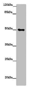
![Various whole cell extracts (30 μg) were separated by 10% SDS-PAGE, and the membrane was blotted with gamma Tubulin antibody [HL1175] (GTX636480) diluted at 1:10000. The HRP-conjugated anti-rabbit IgG antibody (GTX213110-01) was used to detect the primary antibody.](https://www.genetex.com/upload/website/prouct_img/normal/GTX636480/GTX636480_44480_20211126_WB_M_R_w_23061202_303.webp "Various whole cell extracts (30 μg) were separated by 10% SDS-PAGE, and the membrane was blotted with gamma Tubulin antibody [HL1175] (GTX636480) diluted at 1:10000. The HRP-conjugated anti-rabbit IgG antibody (GTX213110-01) was used to detect the primary antibody.")
![Whole cell extract (30 μg) was separated by 7.5% SDS-PAGE, and the membrane was blotted with gamma Tubulin antibody [HL1175] (GTX636480) diluted at 1:1000. The HRP-conjugated anti-rabbit IgG antibody (GTX213110-01) was used to detect the primary antibody.](https://www.genetex.com/upload/website/prouct_img/normal/GTX636480/GTX636480_44480_20230811_WB_Drosophila_23081619_932.webp "Whole cell extract (30 μg) was separated by 7.5% SDS-PAGE, and the membrane was blotted with gamma Tubulin antibody [HL1175] (GTX636480) diluted at 1:1000. The HRP-conjugated anti-rabbit IgG antibody (GTX213110-01) was used to detect the primary antibody.")
![Various whole cell extracts (30 μg) were separated by 10% SDS-PAGE, and the membrane was blotted with gamma Tubulin antibody [HL1175] (GTX636480) diluted at 1:1000. The HRP-conjugated anti-rabbit IgG antibody (GTX213110-01) was used to detect the primary antibody.](https://www.genetex.com/upload/website/prouct_img/normal/GTX636480/GTX636480_44480_20231110_WB_multiple_species_23111422_432.webp "Various whole cell extracts (30 μg) were separated by 10% SDS-PAGE, and the membrane was blotted with gamma Tubulin antibody [HL1175] (GTX636480) diluted at 1:1000. The HRP-conjugated anti-rabbit IgG antibody (GTX213110-01) was used to detect the primary antibody.")
Various whole cell extracts (30 μg) were separated by 10% SDS-PAGE, and the membrane was blotted with gamma Tubulin antibody [HL1175] (GTX636480) diluted at 1:5000. The HRP-conjugated anti-rabbit IgG antibody (GTX213110-01) was used to detect the primary antibody.
gamma Tubulin antibody [HL1175]
GTX636480
ApplicationsImmunoFluorescence, Western Blot, ImmunoCytoChemistry, ImmunoHistoChemistry, ImmunoHistoChemistry Frozen
Product group Antibodies
ReactivityCanine, Drosophila, Feline, Hamster, Human, Monkey, Mouse, Rabbit, Rat, Zebra Fish
TargetTUBG1
Overview
- SupplierGeneTex
- Product Namegamma Tubulin antibody [HL1175]
- Delivery Days Customer9
- Application Supplier NoteWB: 1:500-1:3000. *Optimal dilutions/concentrations should be determined by the researcher.Not tested in other applications.
- ApplicationsImmunoFluorescence, Western Blot, ImmunoCytoChemistry, ImmunoHistoChemistry, ImmunoHistoChemistry Frozen
- CertificationResearch Use Only
- ClonalityMonoclonal
- Clone IDHL1175
- Concentration1 mg/ml
- ConjugateUnconjugated
- Gene ID7283
- Target nameTUBG1
- Target descriptiontubulin gamma 1
- Target synonymsCDCBM4, GCP-1, TUBG, TUBGCP1, tubulin gamma-1 chain, gamma-tubulin complex component 1, tubulin, gamma polypeptide
- HostRabbit
- IsotypeIgG
- Protein IDP23258
- Protein NameTubulin gamma-1 chain
- ReactivityCanine, Drosophila, Feline, Hamster, Human, Monkey, Mouse, Rabbit, Rat, Zebra Fish
- Storage Instruction-20°C or -80°C,2°C to 8°C
- UNSPSC41116161
Datasheet
Related products
Product group Antibodies
TUBG1 AntibodyCSB-PA025334LA01HU
ApplicationsWestern Blot, ELISA, ImmunoHistoChemistry
ReactivityHuman
TargetTUBG1
- SizePrice
Product group Antibodies
TUBG1 Polyclonal AntibodyCAC14553
ApplicationsWestern Blot, ELISA, ImmunoHistoChemistry
TargetTUBG1
- SizePrice
Product group Antibodies
Anti-TUBG1 Antibody144-06215
ApplicationsWestern Blot
ReactivityHuman, Mouse, Rat
TargetTUBG1
- SizePrice

Product group Antibodies
Anti-TUBG1/2 Antibody Picoband(r)A06313-2-CARRIER-FREE
ApplicationsWestern Blot, ELISA
ReactivityHuman, Mouse, Rat
TargetTUBG1
- SizePrice
Product group Antibodies

TUBG1 / Tubulin Gamma 1 AntibodyLS-C766763
ApplicationsImmunoHistoChemistry
ReactivityMouse
TargetTUBG1
- SizePrice
![WB analysis of SH-SY5Y cells overexpressing TagRFP-tagged or GST-tagged human γ-tubulin 1 (γ-Tb1) or γ-tubulin 2 (γ-Tb2) using GTX79860 gamma Tubulin antibody [TU-32].](https://www.genetex.com/upload/website/prouct_img/normal/GTX79860/GTX79860_20191028_WB_1_w_23061322_913.webp)
Product group Antibodies
gamma Tubulin antibody [TU-32]GTX79860
ApplicationsImmunoFluorescence, Western Blot, ImmunoCytoChemistry
ReactivityHuman, Mouse, Plant, Porcine, Protozoa, Rat
TargetTUBG1
- SizePrice
![ICC/IF analysis of mouse fibroblasts using GTX79861 gamma Tubulin antibody [TU-30]. Red : Primary antibody Blue : DAPI](https://www.genetex.com/upload/website/prouct_img/normal/GTX79861/GTX79861_20191028_ICC-IF_2_w_23061322_442.webp)
Product group Antibodies
gamma Tubulin antibody [TU-30]GTX79861
ApplicationsImmunoFluorescence, Western Blot, ImmunoCytoChemistry
ReactivityBovine, Chicken, Human, Mouse, Plant, Porcine, Protozoa, Rat
TargetTUBG1
- SizePrice
![ICC/IF analysis of HeLa cells using GTX11316 gamma Tubulin antibody [GTU-88] at 1:5,000. Cells were fixed and permeabilized with methanol followed by acetone.](https://www.genetex.com/upload/website/prouct_img/normal/GTX11316/GTX11316_20170605_ICCIF_w_23060500_609.webp)
Product group Antibodies
References
gamma Tubulin antibody [GTU-88]GTX11316
ApplicationsImmunoFluorescence, Western Blot, ELISA, ImmunoCytoChemistry
ReactivityCanine, Chicken, Drosophila, Hamster, Human, Monkey, Mouse, Rat, Xenopus
TargetTUBG1
- SizePrice