

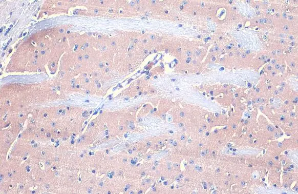
Gephyrin antibody [N2C1], Internal detects Gephyrin protein at cytoplasm by immunohistochemical analysis. Sample: Paraffin-embedded mouse brain. Gephyrin stained by Gephyrin antibody [N2C1], Internal (GTX109734) diluted at 1:500. Antigen Retrieval: Citrate buffer, pH 6.0, 15 min
![Whole cell extract (30 μg) was separated by 7.5% SDS-PAGE, and the membrane was blotted with Gephyrin antibody [N2C1], Internal (GTX109734) diluted at 1:3000. The HRP-conjugated anti-rabbit IgG antibody (GTX213110-01) was used to detect the primary antibody.](https://www.genetex.com/upload/website/prouct_img/normal/GTX109734/GTX109734_44685_20220520_WB_22092622_345.webp "Whole cell extract (30 μg) was separated by 7.5% SDS-PAGE, and the membrane was blotted with Gephyrin antibody [N2C1], Internal (GTX109734) diluted at 1:3000. The HRP-conjugated anti-rabbit IgG antibody (GTX213110-01) was used to detect the primary antibody.")
 A: NIH-3T3 B: JC 7.5% SDS PAGE GTX109734 diluted at 1:1000")
![Immunoprecipitation of Gephyrin protein from A431 whole cell extracts using 5 μg of Gephyrin antibody [N2C1], Internal (GTX109734). Western blot analysis was performed using Gephyrin antibody [N2C1], Internal (GTX109734). EasyBlot anti-Rabbit IgG (GTX221666-01) was used as a secondary reagent.](https://www.genetex.com/upload/website/prouct_img/normal/GTX109734/GTX109734_40884_20150413_IP_w_23060500_773.webp "Immunoprecipitation of Gephyrin protein from A431 whole cell extracts using 5 μg of Gephyrin antibody [N2C1], Internal (GTX109734). Western blot analysis was performed using Gephyrin antibody [N2C1], Internal (GTX109734). EasyBlot anti-Rabbit IgG (GTX221666-01) was used as a secondary reagent.")
![Gephyrin antibody [N2C1], Internal detects Gephyrin protein by immunofluorescent analysis. Sample: DIV10 rat E18 primary hippocampal neurons were fixed in 4% paraformaldehyde at RT for 15 min. Green: Gephyrin protein stained by Gephyrin antibody [N2C1], Internal (GTX109734) diluted at 1:500. Red: beta Tubulin 3/ Tuj1, stained by beta Tubulin 3/ Tuj1 antibody [GT1338] (GTX631831) diluted at 1:500. Blue: Fluoroshield with DAPI (GTX30920).](https://www.genetex.com/upload/website/prouct_img/normal/GTX109734/GTX109734_40884_20170824_IFA_w_23060500_533.webp "Gephyrin antibody [N2C1], Internal detects Gephyrin protein by immunofluorescent analysis. Sample: DIV10 rat E18 primary hippocampal neurons were fixed in 4% paraformaldehyde at RT for 15 min. Green: Gephyrin protein stained by Gephyrin antibody [N2C1], Internal (GTX109734) diluted at 1:500. Red: beta Tubulin 3/ Tuj1, stained by beta Tubulin 3/ Tuj1 antibody [GT1338] (GTX631831) diluted at 1:500. Blue: Fluoroshield with DAPI (GTX30920).")
![Human tissue extract (30 μg) was separated by 7.5% SDS-PAGE, and the membrane was blotted with Gephyrin antibody [N2C1], Internal (GTX109734) diluted at 1:2000. The HRP-conjugated anti-rabbit IgG antibody (GTX213110-01) was used to detect the primary antibody.](https://www.genetex.com/upload/website/prouct_img/normal/GTX109734/GTX109734_43622_20190712_WB_brain_w_23060500_826.webp "Human tissue extract (30 μg) was separated by 7.5% SDS-PAGE, and the membrane was blotted with Gephyrin antibody [N2C1], Internal (GTX109734) diluted at 1:2000. The HRP-conjugated anti-rabbit IgG antibody (GTX213110-01) was used to detect the primary antibody.")
![Gephyrin antibody [N2C1], Internal detects Gephyrin protein expression by immunohistochemical analysis. Sample: Frozen sectioned E13.5 Rat brain. Green: Gephyrin protein stained by Gephyrin antibody [N2C1], Internal (GTX109734) diluted at 1:250. Red: beta Tubulin 3/ TUJ1, a mature neuron marker, stained by beta Tubulin 3/ TUJ1 antibody [GT11710] (GTX631836) diluted at 1:500. Blue: Fluoroshield with DAPI (GTX30920).](https://www.genetex.com/upload/website/prouct_img/normal/GTX109734/GTX109734_40884_20161005_IHC-Fr_R_w_23060500_728.webp "Gephyrin antibody [N2C1], Internal detects Gephyrin protein expression by immunohistochemical analysis. Sample: Frozen sectioned E13.5 Rat brain. Green: Gephyrin protein stained by Gephyrin antibody [N2C1], Internal (GTX109734) diluted at 1:250. Red: beta Tubulin 3/ TUJ1, a mature neuron marker, stained by beta Tubulin 3/ TUJ1 antibody [GT11710] (GTX631836) diluted at 1:500. Blue: Fluoroshield with DAPI (GTX30920).")
![Various tissue extracts (50 μg) were separated by 7.5% SDS-PAGE, and the membrane was blotted with Gephyrin antibody [N2C1], Internal (GTX109734) diluted at 1:2000. The HRP-conjugated anti-rabbit IgG antibody (GTX213110-01) was used to detect the primary antibody.](https://www.genetex.com/upload/website/prouct_img/normal/GTX109734/GTX109734_44741_20220722_WB_M_R_23112922_961.webp "Various tissue extracts (50 μg) were separated by 7.5% SDS-PAGE, and the membrane was blotted with Gephyrin antibody [N2C1], Internal (GTX109734) diluted at 1:2000. The HRP-conjugated anti-rabbit IgG antibody (GTX213110-01) was used to detect the primary antibody.")
Gephyrin antibody [N2C1], Internal detects Gephyrin protein at cytoplasm by immunohistochemical analysis. Sample: Paraffin-embedded mouse brain. Gephyrin stained by Gephyrin antibody [N2C1], Internal (GTX109734) diluted at 1:500. Antigen Retrieval: Citrate buffer, pH 6.0, 15 min
Gephyrin antibody [N2C1], Internal
GTX109734
ApplicationsImmunoFluorescence, ImmunoPrecipitation, Western Blot, ImmunoCytoChemistry, ImmunoHistoChemistry, ImmunoHistoChemistry Frozen, ImmunoHistoChemistry Paraffin
Product group Antibodies
ReactivityHuman, Mouse, Rat
TargetGPHN
Overview
- SupplierGeneTex
- Product NameGephyrin antibody [N2C1], Internal
- Delivery Days Customer9
- Application Supplier NoteWB: 1:500-1:10000. ICC/IF: 1:100-1:1000. IHC-P: 1:100-1:1000. IHC-Fr: 1:100-1:1000. IP: 1:100-1:500. *Optimal dilutions/concentrations should be determined by the researcher.Not tested in other applications.
- ApplicationsImmunoFluorescence, ImmunoPrecipitation, Western Blot, ImmunoCytoChemistry, ImmunoHistoChemistry, ImmunoHistoChemistry Frozen, ImmunoHistoChemistry Paraffin
- CertificationResearch Use Only
- ClonalityPolyclonal
- Concentration0.59 mg/ml
- ConjugateUnconjugated
- Gene ID10243
- Target nameGPHN
- Target descriptiongephyrin
- Target synonymsGEPH, GPH, GPHRYN, HKPX1, MOCODC, gephyrin
- HostRabbit
- IsotypeIgG
- Protein IDQ9NQX3
- Protein NameGephyrin
- Scientific DescriptionThis gene encodes a neuronal assembly protein that anchors inhibitory neurotransmitter receptors to the postsynaptic cytoskeleton via high affinity binding to a receptor subunit domain and tubulin dimers. In nonneuronal tissues, the encoded protein is also required for molybdenum cofactor biosynthesis. Mutations in this gene may be associated with the neurological condition hyperplexia and also lead to molybdenum cofactor deficiency. Numerous alternatively spliced transcript variants encoding different isoforms have been described; however, the full-length nature of all transcript variants is not currently known. [provided by RefSeq]
- ReactivityHuman, Mouse, Rat
- Storage Instruction-20°C or -80°C,2°C to 8°C
- UNSPSC41116161
Datasheet
Related products
Product group Antibodies
Anti-GPHN AntibodyA46933
ApplicationsImmunoHistoChemistry
ReactivityHuman, Mouse, Rat
- SizePrice
Product group Antibodies

Anti-Gephyrin [GC1]Ab02267-10.0
ApplicationsImmunoFluorescence, Western Blot, ELISA, ImmunoHistoChemistry
ReactivityHuman
TargetGPHN
- SizePrice

Product group Antibodies
Anti-Gephyrin/GPHN Antibody Picoband(r)A04560-1-CARRIER-FREE
ApplicationsImmunoFluorescence, Western Blot, ELISA, ImmunoCytoChemistry, ImmunoHistoChemistry
ReactivityHuman, Monkey, Mouse, Rat
TargetGPHN
- SizePrice
Product group Antibodies
Anti-GPHN Antibody144-08572
ApplicationsImmunoFluorescence, Western Blot, ImmunoHistoChemistry
ReactivityHuman, Mouse, Rat
TargetGPHN
- SizePrice
Product group Antibodies
Gephyrin Recombinant Antibody, AbBy Fluor-647 ConjugatedBSM-61891R-BF647
ApplicationsImmunoFluorescence, Western Blot
ReactivityHuman, Mouse, Rat
TargetGPHN
- SizePrice
Product group Antibodies
GPHN AntibodyCSB-PA009716LA01HU
ApplicationsELISA, ImmunoHistoChemistry
ReactivityHuman
TargetGPHN
- SizePrice
Product group Antibodies

Gephyrin AntibodyLS-C403177
ApplicationsELISA, ImmunoHistoChemistry
ReactivityHuman, Mouse, Rat
TargetGPHN
- SizePrice

Product group Antibodies
Anti-GPHN AntibodyHPA024694
ApplicationsWestern Blot, ImmunoHistoChemistry
ReactivityHuman
TargetGPHN
- SizePrice
![Various whole cell extracts (30 μg) were separated by 7.5% SDS-PAGE, and the membrane was blotted with Gephyrin antibody [HL3712] (GTX641902) diluted at 1:1000. The HRP-conjugated anti-rabbit IgG antibody (GTX213110-01) was used to detect the primary antibody. Corresponding RNA expression data for the same cell lines are based on Human Protein Atlas program.](https://www.genetex.com/upload/website/prouct_img/normal/GTX641902/GTX641902_T-45663_20250207_WB_TPM_watermark_25021320_909.webp)
Product group Antibodies
Gephyrin antibody [HL3712]GTX641902
ApplicationsWestern Blot, ImmunoHistoChemistry, ImmunoHistoChemistry Paraffin
ReactivityHuman, Mouse, Rat
TargetGPHN
- SizePrice