

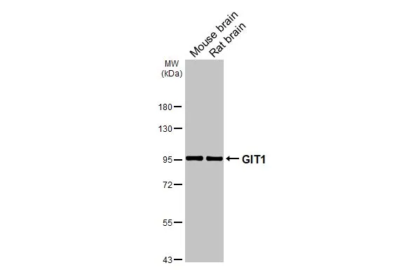
Various tissue extracts (50 μg) were separated by 7.5% SDS-PAGE, and the membrane was blotted with GIT1 antibody [GT87] (GTX641942) diluted at 1:1000. The HRP-conjugated anti-mouse IgG antibody (GTX213111-01) was used to detect the primary antibody.
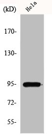
![Various whole cell extracts (30 μg) were separated by 7.5% SDS-PAGE, and the membrane was blotted with GIT1 antibody [GT87] (GTX641942) diluted at 1:1000. The HRP-conjugated anti-mouse IgG antibody (GTX213111-01) was used to detect the primary antibody. Corresponding RNA expression data for the same cell lines are based on Human Protein Atlas program.](https://www.genetex.com/upload/website/prouct_img/normal/GTX641942/GTX641942_45663_20250124_WB_TPM_watermark_25020422_315.webp "Various whole cell extracts (30 μg) were separated by 7.5% SDS-PAGE, and the membrane was blotted with GIT1 antibody [GT87] (GTX641942) diluted at 1:1000. The HRP-conjugated anti-mouse IgG antibody (GTX213111-01) was used to detect the primary antibody. Corresponding RNA expression data for the same cell lines are based on Human Protein Atlas program.")
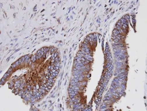
![GIT1 antibody [GT87] detects GIT1 protein by immunohistochemical analysis. Sample: Paraffin-embedded rat cerebellum. GIT1 stained by GIT1 antibody [GT87] (GTX641942) diluted at 1:200. Antigen Retrieval: Citrate buffer, pH 6.0, 15 min](https://www.genetex.com/upload/website/prouct_img/normal/GTX641942/GTX641942_45663_20250606_IHC-P_R_25061300_697.webp "GIT1 antibody [GT87] detects GIT1 protein by immunohistochemical analysis. Sample: Paraffin-embedded rat cerebellum. GIT1 stained by GIT1 antibody [GT87] (GTX641942) diluted at 1:200. Antigen Retrieval: Citrate buffer, pH 6.0, 15 min")
![GIT1 antibody [GT87] detects GIT1 protein by immunohistochemical analysis. Sample: Paraffin-embedded mouse hippocampus. GIT1 stained by GIT1 antibody [GT87] (GTX641942) diluted at 1:100. Antigen Retrieval: Citrate buffer, pH 6.0, 15 min](https://www.genetex.com/upload/website/prouct_img/normal/GTX641942/GTX641942_45663_20250523_IHC-P_M_2_25061300_900.webp "GIT1 antibody [GT87] detects GIT1 protein by immunohistochemical analysis. Sample: Paraffin-embedded mouse hippocampus. GIT1 stained by GIT1 antibody [GT87] (GTX641942) diluted at 1:100. Antigen Retrieval: Citrate buffer, pH 6.0, 15 min")
![GIT1 antibody [GT87] detects GIT1 protein by immunohistochemical analysis. Sample: Paraffin-embedded mouse hippocampus. GIT1 stained by GIT1 antibody [GT87] (GTX641942) diluted at 1:100. Antigen Retrieval: Citrate buffer, pH 6.0, 15 min](https://www.genetex.com/upload/website/prouct_img/normal/GTX641942/GTX641942_45663_20250523_IHC-P_M_1_25061300_838.webp "GIT1 antibody [GT87] detects GIT1 protein by immunohistochemical analysis. Sample: Paraffin-embedded mouse hippocampus. GIT1 stained by GIT1 antibody [GT87] (GTX641942) diluted at 1:100. Antigen Retrieval: Citrate buffer, pH 6.0, 15 min")
![GIT1 antibody [GT87] detects GIT1 protein by immunohistochemical analysis. Sample: Paraffin-embedded mouse tissues. GIT1 stained by GIT1 antibody [GT87] (GTX641942) diluted at 1:100. Antigen Retrieval: Citrate buffer, pH 6.0, 15 min](https://www.genetex.com/upload/website/prouct_img/normal/GTX641942/GTX641942_45663_20250523_IHC-P_M_Multiple_RPKM_25061300_251.webp "GIT1 antibody [GT87] detects GIT1 protein by immunohistochemical analysis. Sample: Paraffin-embedded mouse tissues. GIT1 stained by GIT1 antibody [GT87] (GTX641942) diluted at 1:100. Antigen Retrieval: Citrate buffer, pH 6.0, 15 min")
![GIT1 antibody [GT87] detects GIT1 protein by immunofluorescent analysis. Sample: U87-MG cells were fixed in 4% paraformaldehyde at RT for 15 min. Green: GIT1 stained by GIT1 antibody [GT87] (GTX641942) diluted at 1:500. Red: alpha Tubulin, a cytoskeleton marker, stained by alpha Tubulin antibody [HL2162] (GTX638140) diluted at 1:500. Blue: Fluoroshield with DAPI (GTX30920).](https://www.genetex.com/upload/website/prouct_img/normal/GTX641942/GTX641942_45663_20250613_ICC_IF_25061901_356.webp "GIT1 antibody [GT87] detects GIT1 protein by immunofluorescent analysis. Sample: U87-MG cells were fixed in 4% paraformaldehyde at RT for 15 min. Green: GIT1 stained by GIT1 antibody [GT87] (GTX641942) diluted at 1:500. Red: alpha Tubulin, a cytoskeleton marker, stained by alpha Tubulin antibody [HL2162] (GTX638140) diluted at 1:500. Blue: Fluoroshield with DAPI (GTX30920).")
Various tissue extracts (50 μg) were separated by 7.5% SDS-PAGE, and the membrane was blotted with GIT1 antibody [GT87] (GTX641942) diluted at 1:1000. The HRP-conjugated anti-mouse IgG antibody (GTX213111-01) was used to detect the primary antibody.
GIT1 antibody [GT87]
GTX641942
ApplicationsImmunoFluorescence, Western Blot, ImmunoCytoChemistry, ImmunoHistoChemistry, ImmunoHistoChemistry Paraffin
Product group Antibodies
ReactivityHuman, Mouse, Rat
TargetGIT1
Overview
- SupplierGeneTex
- Product NameGIT1 antibody [GT87]
- Delivery Days Customer9
- Application Supplier NoteWB: 1:500-1:3000. *Optimal dilutions/concentrations should be determined by the researcher.Not tested in other applications.
- ApplicationsImmunoFluorescence, Western Blot, ImmunoCytoChemistry, ImmunoHistoChemistry, ImmunoHistoChemistry Paraffin
- CertificationResearch Use Only
- ClonalityMonoclonal
- Clone IDGT87
- Concentration1 mg/ml
- ConjugateUnconjugated
- Gene ID28964
- Target nameGIT1
- Target descriptionGIT ArfGAP 1
- Target synonymsp95-APP1, ARF GTPase-activating protein GIT1, ARF GAP GIT1, CAT-1, CAT1, G protein-coupled receptor kinase interacting ArfGAP 1, G protein-coupled receptor kinase-interactor 1, GRK-interacting protein 1, cool-associated and tyrosine-phosphorylated protein 1
- HostMouse
- IsotypeIgG2b
- Protein IDQ9Y2X7
- Protein NameARF GTPase-activating protein GIT1
- Scientific DescriptionEnables gamma-tubulin binding activity. Involved in positive regulation of microtubule nucleation and regulation of cytokinesis. Located in several cellular components, including focal adhesion; microtubule cytoskeleton; and mitochondrion. Implicated in attention deficit hyperactivity disorder. Biomarker of Huntingtons disease. [provided by Alliance of Genome Resources, Jan 2025]
- ReactivityHuman, Mouse, Rat
- Storage Instruction-20°C or -80°C,2°C to 8°C
- UNSPSC41116161
Related products
Product group Antibodies
GIT1 AntibodyCSB-PA002688
ApplicationsWestern Blot, ELISA, ImmunoHistoChemistry
ReactivityHuman, Mouse, Rat
TargetGIT1
- SizePrice

Product group Antibodies
Anti-GIT1 Antibody Picoband(r)A02140-3-CARRIER-FREE
ApplicationsFlow Cytometry, ImmunoFluorescence, Western Blot, ELISA, ImmunoCytoChemistry, ImmunoHistoChemistry
ReactivityHuman, Mouse, Rat
TargetGIT1
- SizePrice
Product group Antibodies
Anti-GIT1 Antibody144-61053
ApplicationsWestern Blot
ReactivityHuman, Mouse, Rat
TargetGIT1
- SizePrice

Product group Antibodies
Anti-GIT1 AntibodyA96195
ApplicationsWestern Blot, ELISA, ImmunoHistoChemistry
ReactivityHuman, Mouse, Rat
- SizePrice
Product group Antibodies

GIT1 AntibodyLS-C750405
ApplicationsWestern Blot
ReactivityHuman, Mouse, Rat
TargetGIT1
- SizePrice

Product group Antibodies
Anti-GIT1 AntibodyHPA004059
ApplicationsWestern Blot, ImmunoCytoChemistry, ImmunoHistoChemistry
ReactivityHuman
TargetGIT1
- SizePrice
Product group Antibodies
GIT1 antibody [N3C2], InternalGTX105824
ApplicationsImmunoFluorescence, Western Blot, ImmunoCytoChemistry, ImmunoHistoChemistry, ImmunoHistoChemistry Paraffin
ReactivityHuman, Mouse, Rat
TargetGIT1
- SizePrice
Product group Antibodies
GIT1 Recombinant AntibodyBSM-61866R
ApplicationsFlow Cytometry, Western Blot
ReactivityHuman, Mouse, Rat
TargetGIT1
- SizePrice