

GLUT-1 (Tumor Progression and Mesothelioma Marker)(rGLUT1/2476), CF594 conjugate, 0.1mg/mL [26628-22-8]
BNC943795
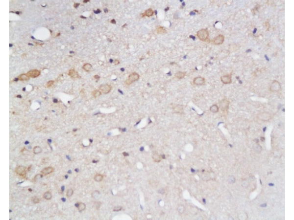
ApplicationsImmunoHistoChemistry, ImmunoHistoChemistry Paraffin
Product group Antibodies
ReactivityHuman
TargetSLC2A1
Overview
- SupplierBiotium
- Product NameGLUT-1 (Tumor Progression and Mesothelioma Marker)(rGLUT1/2476), CF594 conjugate, 0.1mg/mL [26628-22-8]
- Delivery Days Customer9
- ApplicationsImmunoHistoChemistry, ImmunoHistoChemistry Paraffin
- CAS Number26628-22-8
- CertificationResearch Use Only
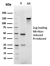
- ClonalityMonoclonal
- Clone IDrGLUT1/2476
- Concentration0.1 mg/ml
- ConjugateOther Conjugate
- Gene ID6513
- Target nameSLC2A1
- Target descriptionsolute carrier family 2 member 1
- Target synonymsCSE, DYT17, DYT18, DYT9, EIG12, GLUT, GLUT-1, GLUT1, GLUT1DS, HTLVR, PED, SDCHCN, solute carrier family 2, facilitated glucose transporter member 1, choreoathetosis/spasticity, episodic (paroxysmal choreoathetosis/spasticity), dystonia gene 18, dystonia gene 9, glucose transporter type 1, erythrocyte/brain, hepG2 glucose transporter, human T-cell leukemia virus (I and II) receptor, receptor for HTLV-1 and HTLV-2, solute carrier family 2 (facilitated glucose transporter), member 1
- HostMouse
- IsotypeIgG1
- Protein IDP11166
- Protein NameSolute carrier family 2, facilitated glucose transporter member 1
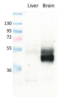
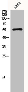
- Scientific DescriptionRecognizes a protein of 55 kDa, which is identified as GLUT-1. Glucose transporters are integral membrane glycoproteins involved in transporting glucose into most cells. There are many types of glucose transport carrier proteins, designated as Glut-1 to Glut-12. Glut-1 is a major glucose transporter in the mammalian blood-brain barrier. It is expressed in high density on the membranes of human erythrocytes and the brain capillaries that comprise the blood-brain barrier. Glut-1 is expressed at variable levels in many human tissues. Overexpression of Glut-1 has been linked to tumor progression or poor survival of patients with carcinomas of the colon, breast, cervical, lung, bladder and mesothelioma. Glut-1 is a sensitive and specific marker for the differentiation of malignant mesothelioma (positive) from reactive mesothelium (negative). Primary antibodies are available purified, or with a selection of fluorescent CF® Dyes and other labels. CF® Dyes offer exceptional brightness and photostability. Note: Conjugates of blue fluorescent dyes like CF®405S and CF®405M are not recommended for detecting low abundance targets, because blue dyes have lower fluorescence and can give higher non-specific background than other dye colors.
- SourceAnimal
- ReactivityHuman
- Storage Instruction2°C to 8°C,RT
- UNSPSC41116161
MSDS
Related products
Product group Antibodies
Anti-GLUT1 AntibodyA95873
ApplicationsWestern Blot, ELISA, ImmunoHistoChemistry
ReactivityHuman, Mouse, Rat
- SizePrice
Product group Antibodies
anti-GLUT1, pAb (IN116)AG-25B-0040
ApplicationsImmunoPrecipitation, Western Blot, ImmunoHistoChemistry
ReactivityHuman, Mouse, Rat
TargetSLC2A1
- SizePrice
Product group Antibodies
Anti-SLC2A1 Antibody144-06982
ApplicationsWestern Blot
ReactivityAvian, Human, Mouse, Rat
TargetSLC2A1
- SizePrice
Product group Antibodies
References
GLUT1 Polyclonal Antibodybs-0472R
ApplicationsFlow Cytometry, ImmunoFluorescence, Western Blot, ELISA, ImmunoCytoChemistry, ImmunoHistoChemistry, ImmunoHistoChemistry Frozen, ImmunoHistoChemistry Paraffin
ReactivityBovine, Canine, Chicken, Human, Mouse, Porcine, Rat, Sheep
TargetSLC2A1
- SizePrice
Product group Antibodies
SLC2A1 AntibodyCSB-PA002728
ApplicationsWestern Blot, ELISA, ImmunoHistoChemistry
ReactivityHuman, Mouse, Rat
TargetSLC2A1
- SizePrice
Product group Antibodies
Slc2A1 Polyclonal AntibodyCAC10560
ApplicationsWestern Blot, ELISA, ImmunoHistoChemistry
ReactivityMouse
TargetSLC2A1
- SizePrice
![IHC-P analysis of human cerebrum (grey matter) tissue using GTX04469 GluT1 antibody [MSVA-401R] HistoMAX?. A particularly strong GluT1 staining of endothelial cells is seen in the brain.](https://www.genetex.com/upload/website/prouct_img/normal/GTX04469/GTX04469_20230728_IHC-P_56_23072722_577.webp)
Product group Antibodies
ApplicationsImmunoHistoChemistry, ImmunoHistoChemistry Paraffin
ReactivityHuman
TargetSLC2A1
- SizePrice

Product group Antibodies
Anti-SLC2A1 AntibodyHPA031345
ApplicationsImmunoCytoChemistry, ImmunoHistoChemistry
ReactivityHuman, Mouse
TargetSLC2A1
- SizePrice

Product group Antibodies
Anti-Glucose Transporter GLUT1/SLC2A1 Antibody Picoband(r)PB9435-CARRIER-FREE
ApplicationsFlow Cytometry, ImmunoFluorescence, Western Blot, ImmunoCytoChemistry, ImmunoHistoChemistry, ImmunoHistoChemistry Frozen
ReactivityHuman, Mouse, Rat
TargetSLC2A1
- SizePrice