


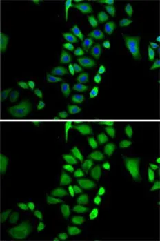
Glyoxalase I antibody [GT266] detects Glyoxalase I protein at cytoplasm by immunofluorescent analysis. Sample: HeLa cells were fixed in 4% paraformaldehyde at RT for 15 min. Green: Glyoxalase I stained by Glyoxalase I antibody [GT266] (GTX628889) diluted at 1:100. Blue: Hoechst 33342 staining. Scale bar= 10 μm.
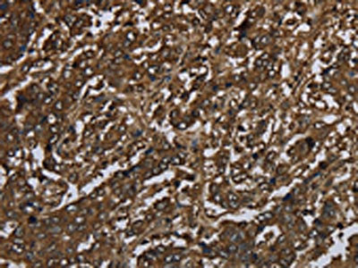
![Glyoxalase I antibody [GT266] detects Glyoxalase I protein at cytoplasm by immunohistochemical analysis. Sample: Paraffin-embedded human ovarian cancer. Glyoxalase I stained by Glyoxalase I antibody [GT266] (GTX628889) diluted at 1:200. Antigen Retrieval: Citrate buffer, pH 6.0, 15 min](https://www.genetex.com/upload/website/prouct_img/normal/GTX628889/GTX628889_41295_20180622_IHC-P_w_23061202_696.webp "Glyoxalase I antibody [GT266] detects Glyoxalase I protein at cytoplasm by immunohistochemical analysis. Sample: Paraffin-embedded human ovarian cancer. Glyoxalase I stained by Glyoxalase I antibody [GT266] (GTX628889) diluted at 1:200. Antigen Retrieval: Citrate buffer, pH 6.0, 15 min")
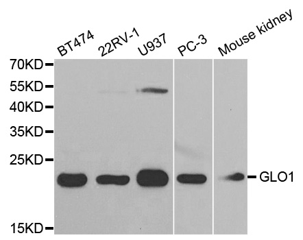
![Glyoxalase I antibody [GT266] detects GLO1 protein by Western blot analysis. A. 30 μg 293T whole cell lysate/extract B. 30 μg HeLa whole cell lysate/extract C. 30 μg HepG2 whole cell lysate/extract D. 30 μg A375 whole cell lysate/extract 12 % SDS-PAGE Glyoxalase I antibody [GT266] (GTX628889) dilution: 1:1000](https://www.genetex.com/upload/website/prouct_img/normal/GTX628889/GTX628889_41295_WB_w_23061202_285.webp "Glyoxalase I antibody [GT266] detects GLO1 protein by Western blot analysis. A. 30 μg 293T whole cell lysate/extract B. 30 μg HeLa whole cell lysate/extract C. 30 μg HepG2 whole cell lysate/extract D. 30 μg A375 whole cell lysate/extract 12 % SDS-PAGE Glyoxalase I antibody [GT266] (GTX628889) dilution: 1:1000")
![Non-transfected (–) and transfected (+) 293T whole cell extracts (30 μg) were separated by 12% SDS-PAGE, and the membrane was blotted with Glyoxalase I antibody [GT266] (GTX628889) diluted at 1:1000. The HRP-conjugated anti-mouse IgG antibody (GTX213111-01) was used to detect the primary antibody.](https://www.genetex.com/upload/website/prouct_img/normal/GTX628889/GTX628889_41295_20200807_WB_shRNA_watermark_w_23061202_809.webp "Non-transfected (–) and transfected (+) 293T whole cell extracts (30 μg) were separated by 12% SDS-PAGE, and the membrane was blotted with Glyoxalase I antibody [GT266] (GTX628889) diluted at 1:1000. The HRP-conjugated anti-mouse IgG antibody (GTX213111-01) was used to detect the primary antibody.")
Glyoxalase I antibody [GT266] detects Glyoxalase I protein at cytoplasm by immunofluorescent analysis. Sample: HeLa cells were fixed in 4% paraformaldehyde at RT for 15 min. Green: Glyoxalase I stained by Glyoxalase I antibody [GT266] (GTX628889) diluted at 1:100. Blue: Hoechst 33342 staining. Scale bar= 10 μm.
Glyoxalase I antibody [GT266]
GTX628889
ApplicationsImmunoFluorescence, Western Blot, ImmunoCytoChemistry, ImmunoHistoChemistry, ImmunoHistoChemistry Paraffin
Product group Antibodies
ReactivityHuman
TargetGLO1
Overview
- SupplierGeneTex
- Product NameGlyoxalase I antibody [GT266]
- Delivery Days Customer9
- Application Supplier NoteWB: 1:500-1:3000. ICC/IF: 1:100-1:1000. IHC-P: 1:100-1:1000. *Optimal dilutions/concentrations should be determined by the researcher.Not tested in other applications.
- ApplicationsImmunoFluorescence, Western Blot, ImmunoCytoChemistry, ImmunoHistoChemistry, ImmunoHistoChemistry Paraffin
- CertificationResearch Use Only
- ClonalityMonoclonal
- Clone IDGT266
- Concentration1 mg/ml
- ConjugateUnconjugated
- Gene ID2739
- Target nameGLO1
- Target descriptionglyoxalase I
- Target synonymsGLOD1, GLYI, HEL-S-74, lactoylglutathione lyase, S-D-lactoylglutathione methylglyoxal lyase, aldoketomutase, epididymis secretory protein Li 74, glx I, glyoxalase domain containing 1, ketone-aldehyde mutase, lactoyl glutathione lyase, methylglyoxalase
- HostMouse
- IsotypeIgG2b
- Protein IDQ04760
- Protein NameLactoylglutathione lyase
- Scientific DescriptionThe enzyme encoded by this gene is responsible for the catalysis and formation of S-lactoyl-glutathione from methylglyoxal condensation and reduced glutatione. Glyoxalase I is linked to HLA and is localized to 6p21.3-p21.1, between HLA and the centromere. [provided by RefSeq]
- ReactivityHuman
- Storage Instruction-20°C or -80°C,2°C to 8°C
- UNSPSC41116161
Datasheet
Related products
Product group Antibodies
Anti-GLO1 AntibodyA30523
ApplicationsWestern Blot, ImmunoHistoChemistry
ReactivityHuman, Mouse, Rat
- SizePrice

Product group Antibodies
Anti-GLO1 Antibody Picoband(r)A01703-1-CARRIER-FREE
ApplicationsImmunoPrecipitation, Western Blot, ELISA, ImmunoHistoChemistry
ReactivityHuman, Mouse, Rat
TargetGLO1
- SizePrice
Product group Antibodies
GLO1 Recombinant Antibody, AbBy Fluor-488 ConjugatedBSM-61735R-BF488
ApplicationsFlow Cytometry, Western Blot
ReactivityHuman, Mouse, Rat
TargetGLO1
- SizePrice
Product group Antibodies
GLO1 AntibodyCSB-PA829442
ApplicationsWestern Blot, ELISA, ImmunoHistoChemistry
ReactivityHuman, Mouse, Rat
TargetGLO1
- SizePrice
Product group Antibodies
Glo1 Polyclonal AntibodyCAC11639
ApplicationsWestern Blot, ELISA
ReactivityPlant
- SizePrice
Product group Antibodies

GLO1 / Glyoxalase I AntibodyLS-C405019
ApplicationsWestern Blot, ELISA, ImmunoHistoChemistry
ReactivityHuman, Mouse, Rat
TargetGLO1
- SizePrice
![IHC-P analysis of human prostate and stomach carcinoma tissue using GTX15747 Glyoxalase I antibody [Glo1a]. Antigen retrieval : heat induced antigen retrieval was performed using 10mM sodium citrate (pH6.0) buffer for 20 minutes Dilution : 1:800](https://www.genetex.com/upload/website/prouct_img/normal/GTX15747/GTX15747_1035_IHC-P_w_23060620_356.webp)
Product group Antibodies
References
Glyoxalase I antibody [Glo1a]GTX15747
ApplicationsImmunoFluorescence, Western Blot, ImmunoCytoChemistry, ImmunoHistoChemistry, ImmunoHistoChemistry Paraffin
ReactivityHuman, Mouse, Primate, Rat
TargetGLO1
- SizePrice

Product group Antibodies
Anti-GLO1 AntibodyHPA059791
ApplicationsWestern Blot, ImmunoCytoChemistry
ReactivityHuman
TargetGLO1
- SizePrice
![Glyoxalase I antibody [N1C3] detects Glyoxalase I protein at cytoplasm by immunohistochemical analysis. Sample: Paraffin-embedded human ovarian cancer. Glyoxalase I stained by Glyoxalase I antibody [N1C3] (GTX105792) diluted at 1:500. Antigen Retrieval: Citrate buffer, pH 6.0, 15 min](https://www.genetex.com/upload/website/prouct_img/normal/GTX105792/GTX105792_43495_20190517_IHC-P_w_23060120_933.webp)
Product group Antibodies
Glyoxalase I antibody [N1C3]GTX105792
ApplicationsImmunoFluorescence, Western Blot, ImmunoCytoChemistry, ImmunoHistoChemistry, ImmunoHistoChemistry Paraffin
ReactivityHuman, Mouse
TargetGLO1
- SizePrice
Product group Antibodies
Glyoxalase I antibodyGTX55643
ApplicationsImmunoFluorescence, Western Blot, ImmunoCytoChemistry, ImmunoHistoChemistry, ImmunoHistoChemistry Paraffin
ReactivityHuman, Mouse
TargetGLO1
- SizePrice