

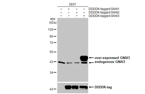
Non-transfected (–) and transfected (+) 293T whole cell extracts (30 μg) were separated by 10% SDS-PAGE, and the membrane was blotted with GNAI3 antibody [HL2096] (GTX638004) diluted at 1:5000. The HRP-conjugated anti-rabbit IgG antibody (GTX213110-01) was used to detect the primary antibody.
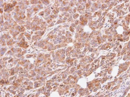
![GNAI3 antibody [HL2096] detects GNAI3 protein by immunohistochemical analysis. Sample: Paraffin-embedded mouse tissues. GNAI3 stained by GNAI3 antibody [HL2096] (GTX638004) diluted at 1:100. Antigen Retrieval: Citrate buffer, pH 6.0, 15 min](https://www.genetex.com/upload/website/prouct_img/normal/GTX638004/GTX638004_T-44900_20230113_IHC-P_multiple_M_23013122_244.webp "GNAI3 antibody [HL2096] detects GNAI3 protein by immunohistochemical analysis. Sample: Paraffin-embedded mouse tissues. GNAI3 stained by GNAI3 antibody [HL2096] (GTX638004) diluted at 1:100. Antigen Retrieval: Citrate buffer, pH 6.0, 15 min")
![Wild-type (WT) and GNAI3 knockout (KO) HeLa cell extracts (30 μg) were separated by 10% SDS-PAGE, and the membrane was blotted with GNAI3 antibody [HL2096] (GTX638004) diluted at 1:1000. The HRP-conjugated anti-rabbit IgG antibody (GTX213110-01) was used to detect the primary antibody.](https://www.genetex.com/upload/website/prouct_img/normal/GTX638004/GTX638004_T-44900_20230120_WB_KO_watermark_23013122_138.webp "Wild-type (WT) and GNAI3 knockout (KO) HeLa cell extracts (30 μg) were separated by 10% SDS-PAGE, and the membrane was blotted with GNAI3 antibody [HL2096] (GTX638004) diluted at 1:1000. The HRP-conjugated anti-rabbit IgG antibody (GTX213110-01) was used to detect the primary antibody.")
![Various tissue extracts (50 μg) were separated by 10% SDS-PAGE, and the membrane was blotted with GNAI3 antibody [HL2096] (GTX638004) diluted at 1:1000. The HRP-conjugated anti-rabbit IgG antibody (GTX213110-01) was used to detect the primary antibody, and the signal was developed with Trident ECL plus-Enhanced.](https://www.genetex.com/upload/website/prouct_img/normal/GTX638004/GTX638004_T-44900_20230120_WB_M_R_23013122_762.webp "Various tissue extracts (50 μg) were separated by 10% SDS-PAGE, and the membrane was blotted with GNAI3 antibody [HL2096] (GTX638004) diluted at 1:1000. The HRP-conjugated anti-rabbit IgG antibody (GTX213110-01) was used to detect the primary antibody, and the signal was developed with Trident ECL plus-Enhanced.")
![GNAI3 antibody [HL2096] detects GNAI3 protein by immunohistochemical analysis. Sample: Paraffin-embedded rat tissues. GNAI3 stained by GNAI3 antibody [HL2096] (GTX638004) diluted at 1:100. Antigen Retrieval: Citrate buffer, pH 6.0, 15 min](https://www.genetex.com/upload/website/prouct_img/normal/GTX638004/GTX638004_T-44900_20230113_IHC-P_multiple_R_23013122_125.webp "GNAI3 antibody [HL2096] detects GNAI3 protein by immunohistochemical analysis. Sample: Paraffin-embedded rat tissues. GNAI3 stained by GNAI3 antibody [HL2096] (GTX638004) diluted at 1:100. Antigen Retrieval: Citrate buffer, pH 6.0, 15 min")
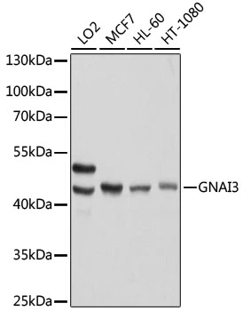
![Various whole cell extracts (30 μg) were separated by 10% SDS-PAGE, and the membrane was blotted with GNAI3 antibody [HL2096] (GTX638004) diluted at 1:1000. The HRP-conjugated anti-rabbit IgG antibody (GTX213110-01) was used to detect the primary antibody.](https://www.genetex.com/upload/website/prouct_img/normal/GTX638004/GTX638004_44977_20230317_WB_23032022_220.webp "Various whole cell extracts (30 μg) were separated by 10% SDS-PAGE, and the membrane was blotted with GNAI3 antibody [HL2096] (GTX638004) diluted at 1:1000. The HRP-conjugated anti-rabbit IgG antibody (GTX213110-01) was used to detect the primary antibody.")
![GNAI3 antibody [HL2096] detects GNAI3 protein by immunofluorescent analysis. Sample: A431 cells were fixed in ice-cold MeOH for 5 min. Green: GNAI3 stained by GNAI3 antibody [HL2096] (GTX638004) diluted at 1:500. Red: alpha Tubulin, a cytoskeleton marker, stained by alpha Tubulin antibody [GT114] (GTX628802) diluted at 1:1000. Blue: Fluoroshield with DAPI (GTX30920).](https://www.genetex.com/upload/website/prouct_img/normal/GTX638004/GTX638004_44977_20240607_ICC_IF_24070822_954.webp "GNAI3 antibody [HL2096] detects GNAI3 protein by immunofluorescent analysis. Sample: A431 cells were fixed in ice-cold MeOH for 5 min. Green: GNAI3 stained by GNAI3 antibody [HL2096] (GTX638004) diluted at 1:500. Red: alpha Tubulin, a cytoskeleton marker, stained by alpha Tubulin antibody [GT114] (GTX628802) diluted at 1:1000. Blue: Fluoroshield with DAPI (GTX30920).")
Non-transfected (–) and transfected (+) 293T whole cell extracts (30 μg) were separated by 10% SDS-PAGE, and the membrane was blotted with GNAI3 antibody [HL2096] (GTX638004) diluted at 1:5000. The HRP-conjugated anti-rabbit IgG antibody (GTX213110-01) was used to detect the primary antibody.
GNAI3 antibody [HL2096]
GTX638004
ApplicationsImmunoFluorescence, Western Blot, ImmunoCytoChemistry, ImmunoHistoChemistry, ImmunoHistoChemistry Paraffin
Product group Antibodies
ReactivityHuman, Mouse, Rat
TargetGNAI3
Overview
- SupplierGeneTex
- Product NameGNAI3 antibody [HL2096]
- Delivery Days Customer9
- Application Supplier NoteWB: 1:500-1:10000. *Optimal dilutions/concentrations should be determined by the researcher.Not tested in other applications.
- ApplicationsImmunoFluorescence, Western Blot, ImmunoCytoChemistry, ImmunoHistoChemistry, ImmunoHistoChemistry Paraffin
- CertificationResearch Use Only
- ClonalityMonoclonal
- Clone IDHL2096
- Concentration1 mg/ml
- ConjugateUnconjugated
- Gene ID2773
- Target nameGNAI3
- Target descriptionG protein subunit alpha i3
- Target synonyms87U6, ARCND1, ARCODS, HG1A, guanine nucleotide-binding protein G(i) subunit alpha-3, g(i) alpha-3, guanine nucleotide binding protein (G protein), alpha inhibiting activity polypeptide 3, guanine nucleotide-binding protein G(i) subunit alpha, guanine nucleotide-binding protein G(k) subunit alpha, heterotrimeric guanine nucleotide-binding protein 1A
- HostRabbit
- IsotypeIgG
- Protein IDP08754
- Protein NameGuanine nucleotide-binding protein G(i) subunit alpha-3
- Scientific DescriptionGuanine nucleotide-binding proteins (G proteins) are involved as modulators or transducers in various transmembrane signaling pathways. G proteins are composed of 3 units: alpha, beta and gamma. This gene encodes an alpha subunit and belongs to the G-alpha family. Mutation in this gene, resulting in a gly40-to-arg substitution, is associated with auriculocondylar syndrome, and shown to affect downstream targets in the G protein-coupled endothelin receptor pathway. [provided by RefSeq, Jun 2012]
- ReactivityHuman, Mouse, Rat
- Storage Instruction-20°C or -80°C,2°C to 8°C
- UNSPSC41116161
Datasheet
Related products
Product group Antibodies
ReactivityHuman
TargetGNAI3
- SizePrice
Product group Antibodies
GNAI3 AntibodyCSB-PA008887
ApplicationsWestern Blot, ELISA
ReactivityHuman, Mouse, Rat
TargetGNAI3
- SizePrice
Product group Antibodies
Anti-GNAI3 AntibodyA97509
ApplicationsWestern Blot, ELISA
ReactivityHuman, Mouse, Rat
- SizePrice
Product group Antibodies

GNAI3 AntibodyLS-C408378
ApplicationsWestern Blot, ImmunoHistoChemistry
ReactivityHuman, Mouse, Rat
TargetGNAI3
- SizePrice
Product group Antibodies
GNAI3 antibody [N2C3]GTX103676
ApplicationsWestern Blot, ImmunoHistoChemistry, ImmunoHistoChemistry Paraffin
ReactivityHuman, Mouse
TargetGNAI3
- SizePrice
Product group Antibodies
GNAI3 antibodyGTX35215
ApplicationsImmunoFluorescence, ImmunoPrecipitation, Western Blot, ImmunoCytoChemistry
ReactivityHuman, Mouse
TargetGNAI3
- SizePrice
Product group Antibodies
Anti-GNAI3 Antibody144-61143
ApplicationsImmunoFluorescence, Western Blot
ReactivityHuman, Mouse, Rat
TargetGNAI3
- SizePrice
Product group Antibodies
GNAI3 Polyclonal AntibodyBS-1429R
ApplicationsImmunoFluorescence, ELISA, ImmunoCytoChemistry, ImmunoHistoChemistry, ImmunoHistoChemistry Frozen, ImmunoHistoChemistry Paraffin
ReactivityBovine, Canine, Chicken, Equine, Human, Mouse, Porcine, Rat
TargetGNAI3
- SizePrice