

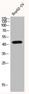
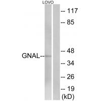
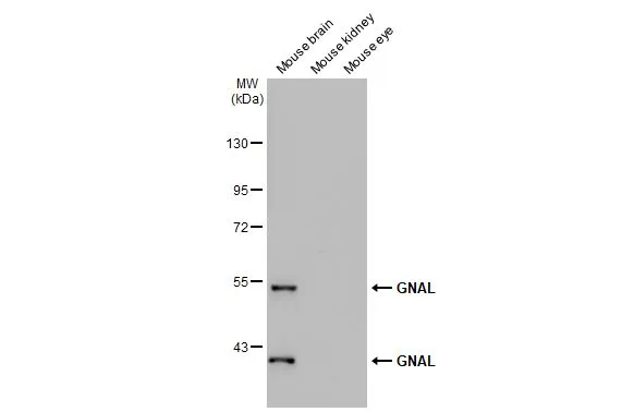
GNAL Polyclonal Antibody
BS-12003R
ApplicationsImmunoFluorescence, Western Blot, ELISA, ImmunoCytoChemistry, ImmunoHistoChemistry, ImmunoHistoChemistry Frozen, ImmunoHistoChemistry Paraffin
Product group Antibodies
ReactivityBovine, Chicken, Human, Mouse, Porcine, Rabbit, Rat, Sheep
Overview
- SupplierBioss
- Product NameGNAL Polyclonal Antibody
- Delivery Days Customer16
- ApplicationsImmunoFluorescence, Western Blot, ELISA, ImmunoCytoChemistry, ImmunoHistoChemistry, ImmunoHistoChemistry Frozen, ImmunoHistoChemistry Paraffin
- Applications SupplierWB(1:300-5000), ELISA(1:500-1000), IHC-P(1:200-400), IHC-F(1:100-500), IF(IHC-P)(1:50-200), IF(IHC-F)(1:50-200), IF(ICC)(1:50-200)
- CertificationResearch Use Only
- ClonalityPolyclonal
- Concentration1 ug/ul
- ConjugateUnconjugated
- HostRabbit
- IsotypeIgG
- ReactivityBovine, Chicken, Human, Mouse, Porcine, Rabbit, Rat, Sheep
- Storage Instruction-20°C
- UNSPSC41116161
Datasheet
Related products
Product group Antibodies
GNAL AntibodyCSB-PA002857
ApplicationsWestern Blot, ELISA, ImmunoHistoChemistry
ReactivityHuman, Mouse, Rat
TargetGNAL
- SizePrice

Product group Antibodies
Anti-GNAL Antibody Picoband(r)A09404-1-CARRIER-FREE
ApplicationsImmunoFluorescence, Western Blot, ELISA, ImmunoCytoChemistry, ImmunoHistoChemistry
ReactivityHuman, Mouse, Rat
TargetGNAL
- SizePrice
Product group Antibodies
Anti-GNAL AntibodyA38635
ApplicationsWestern Blot, ImmunoHistoChemistry
ReactivityHuman, Mouse, Rat
- SizePrice

Product group Antibodies
Anti-GNAL AntibodyHPA051160
ApplicationsImmunoHistoChemistry
ReactivityHuman
TargetGNAL
- SizePrice
Product group Antibodies

GNAL Antibody (Internal)LS-C358758
ApplicationsWestern Blot
ReactivityBovine, Human, Mouse, Rat
TargetGNAL
- SizePrice
Product group Antibodies
GNAL Polyclonal AntibodyCAC13048
ApplicationsWestern Blot, ELISA, ImmunoHistoChemistry
TargetGNAL
- SizePrice
Product group Antibodies
GNAL antibodyGTX103863
ApplicationsImmunoFluorescence, Western Blot, ImmunoCytoChemistry, ImmunoHistoChemistry, ImmunoHistoChemistry Paraffin
ReactivityHuman, Mouse
TargetGNAL
- SizePrice