

Immunohistochemical analysis of paraffin-embedded zebrafish tissue, using GNAO1 antibody (GTX114439) at 1:300 dilution.
 A: adult zebrafish 10% SDS PAGE GTX114439 diluted at 1:5000")
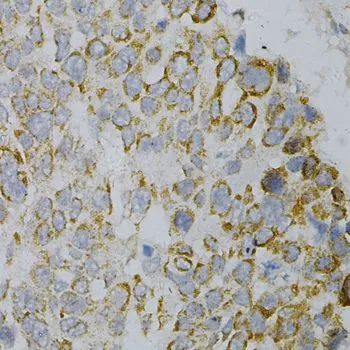
 dilution: 1:500.
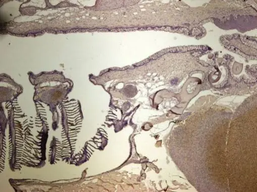
Antigen Retrieval: Trilogy? (EDTA based, pH 8.0) buffer, 15min")
![GNAO1 antibody detects GNAO1 protein by immunohistochemical analysis. Sample: Frozen-sectioned mouse cerebellum. Green: GNAO1 stained by GNAO1 antibody (GTX114439) diluted at 1:250. Red: NF-H, stained by NF-H antibody [GT114] (GTX634289) diluted at 1:500. Blue: Fluoroshield with DAPI (GTX30920).](https://www.genetex.com/upload/website/prouct_img/normal/GTX114439/GTX114439_40198_20180125_IHC-Fr_M_w_23060501_294.webp "GNAO1 antibody detects GNAO1 protein by immunohistochemical analysis. Sample: Frozen-sectioned mouse cerebellum. Green: GNAO1 stained by GNAO1 antibody (GTX114439) diluted at 1:250. Red: NF-H, stained by NF-H antibody [GT114] (GTX634289) diluted at 1:500. Blue: Fluoroshield with DAPI (GTX30920).")
 were separated by 10% SDS-PAGE, and the membrane was blotted with GNAO1 antibody (GTX114439) diluted at 1:10000. The HRP-conjugated anti-rabbit IgG antibody (GTX213110-01) was used to detect the primary antibody.")
 were separated by 10% SDS-PAGE, and the membrane was blotted with GNAO1 antibody (GTX114439) diluted at 1:10000. The HRP-conjugated anti-rabbit IgG antibody (GTX213110-01) was used to detect the primary antibody.")
. Western blot analysis was performed using GNAO1 antibody (GTX114439). EasyBlot anti-Rabbit IgG (GTX221666-01) was used as a secondary reagent.")
 were separated by 10% SDS-PAGE, and the membrane was blotted with GNAO1 antibody (GTX114439) diluted at 1:1000. The HRP-conjugated anti-rabbit IgG antibody (GTX213110-01) was used to detect the primary antibody.")
![GNAO1 antibody detects GNAO1 protein by immunofluorescent analysis. Sample: DIV9 rat E18 primary hippocampal neuron cells were fixed in 4% paraformaldehyde at RT for 15 min. Green: GNAO1 stained by GNAO1 antibody (GTX114439) diluted at 1:500. Red: beta Tubulin 3/ Tuj1, stained by beta Tubulin 3/ Tuj1 antibody [GT1338] (GTX631831) diluted at 1:500. Blue: Fluoroshield with DAPI (GTX30920).](https://www.genetex.com/upload/website/prouct_img/normal/GTX114439/GTX114439_40198_20180131_ICC_IF_R_w_23060501_138.webp "GNAO1 antibody detects GNAO1 protein by immunofluorescent analysis. Sample: DIV9 rat E18 primary hippocampal neuron cells were fixed in 4% paraformaldehyde at RT for 15 min. Green: GNAO1 stained by GNAO1 antibody (GTX114439) diluted at 1:500. Red: beta Tubulin 3/ Tuj1, stained by beta Tubulin 3/ Tuj1 antibody [GT1338] (GTX631831) diluted at 1:500. Blue: Fluoroshield with DAPI (GTX30920).")
 diluted at 1:500.
Antigen Retrieval: Citrate buffer, pH 6.0, 15 min")
Immunohistochemical analysis of paraffin-embedded zebrafish tissue, using GNAO1 antibody (GTX114439) at 1:300 dilution.
GNAO1 antibody
GTX114439
ApplicationsImmunoFluorescence, ImmunoPrecipitation, Western Blot, ImmunoCytoChemistry, ImmunoHistoChemistry, ImmunoHistoChemistry Frozen, ImmunoHistoChemistry Paraffin
Product group Antibodies
ReactivityHuman, Mouse, Rat, Zebra Fish
TargetGNAO1
Overview
- SupplierGeneTex
- Product NameGNAO1 antibody
- Delivery Days Customer9
- Application Supplier NoteWB: 1:500-1:20000. ICC/IF: 1:100-1:1000. IHC-P: 1:100-1:1000. IHC-Fr: 1:100-1:1000. IP: 1:100-1:500. *Optimal dilutions/concentrations should be determined by the researcher.Not tested in other applications.
- ApplicationsImmunoFluorescence, ImmunoPrecipitation, Western Blot, ImmunoCytoChemistry, ImmunoHistoChemistry, ImmunoHistoChemistry Frozen, ImmunoHistoChemistry Paraffin
- CertificationResearch Use Only
- ClonalityPolyclonal
- Concentration1 mg/ml
- ConjugateUnconjugated
- Gene ID2775
- Target nameGNAO1
- Target descriptionG protein subunit alpha o1
- Target synonymsDEE17, EIEE17, G-ALPHA-o, GNAO, HG1G, HLA-DQB1, NEDIM, guanine nucleotide-binding protein G(o) subunit alpha, GO2-q chimeric G-protein, guanine nucleotide binding protein (G protein), alpha activating activity polypeptide O, guanine nucleotide-binding regulatory protein 2, heterotrimeric guanine nucleotide-binding protein 1G
- HostRabbit
- IsotypeIgG
- Protein IDP09471
- Protein NameGuanine nucleotide-binding protein G(o) subunit alpha
- ReactivityHuman, Mouse, Rat, Zebra Fish
- Storage Instruction-20°C or -80°C,2°C to 8°C
- UNSPSC41116161
Datasheet
Related products

Product group Antibodies
Anti-GNAO1 Antibody Picoband(r)A05532-2-CARRIER-FREE
ApplicationsWestern Blot, ELISA
ReactivityHuman, Mouse, Rat
TargetGNAO1
- SizePrice
Product group Antibodies
Anti-GNAO1 AntibodyA282798
ApplicationsWestern Blot, ELISA
ReactivityDrosophila, Mouse
- SizePrice
Product group Antibodies

GNAO1 Antibody (FITC)LS-C318017
ApplicationsWestern Blot, ELISA
ReactivityMouse
TargetGNAO1
- SizePrice

Product group Antibodies
GNAO1 AntibodyCSB-PA009593DSR2HU
ApplicationsELISA, ImmunoHistoChemistry
ReactivityHuman
TargetGNAO1
- SizePrice

Product group Antibodies
Anti-GNAO1 AntibodyHPA040878
ApplicationsWestern Blot, ImmunoHistoChemistry
ReactivityHuman
TargetGNAO1
- SizePrice
Product group Antibodies
GNAO1 antibodyGTX55644
ApplicationsWestern Blot, ImmunoHistoChemistry, ImmunoHistoChemistry Paraffin
ReactivityHuman, Mouse
TargetGNAO1
- SizePrice
Product group Antibodies
Anti-GNAO1 Antibody144-02510
ApplicationsWestern Blot, ImmunoHistoChemistry
ReactivityHuman, Mouse
TargetGNAO1
- SizePrice
Product group Antibodies
GNAO1 Polyclonal AntibodyBS-6962R
ApplicationsImmunoFluorescence, Western Blot, ELISA, ImmunoCytoChemistry, ImmunoHistoChemistry, ImmunoHistoChemistry Frozen, ImmunoHistoChemistry Paraffin
ReactivityBovine, Human, Mouse, Porcine, Rat
TargetGNAO1
- SizePrice