

Goat anti-PINK1
EB07940
ApplicationsFlow Cytometry, Western Blot, ELISA
Product group Antibodies
ReactivityHuman, Rat
TargetPINK1
Overview
- SupplierEverest Biotech
- Product NameGoat anti-PINK1
- Delivery Days Customer5
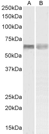
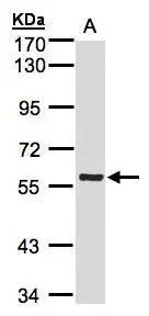
- Application Supplier NoteImmunofluorescence: Strong expression of the protein seen in the mitochondria and endoplasmic reticulum of HeLa cells. Recommended concentration: 10microg/ml. Flow Cytometry: Flow cytometric analysis of MCF7 cells. Recommended concentration: 10ug/ml.
- ApplicationsFlow Cytometry, Western Blot, ELISA
- Applications SupplierPep-ELISA, IF, FC
- CertificationResearch Use Only
- ClonalityPolyclonal
- Concentration0.5 mg/ml
- Gene ID65018
- Target namePINK1
- Target descriptionPTEN induced kinase 1
- Target synonymsBRPK, PARK6, serine/threonine-protein kinase PINK1, mitochondrial, PTEN induced putative kinase 1, PTEN-induced putative kinase protein 1, protein kinase BRPK
- HostGoat
- Scientific DescriptionRefSeq number(s): NP_115785.1. GeneIDs all Nonhuman: 68943 (mouse); 298575 (rat);. Purification: Antigen affinity purified. Names and symbols: PINK1; PTEN induced putative kinase 1 ; BRPK; FLJ27236; PARK6 ; protein kinase BRPK; serine/threonine-protein kinase PINK1
- ReactivityHuman, Rat
- Reactivity SupplierHuman, Rat
- Storage Instruction-20°C
- UNSPSC12352203
Related products
Product group Antibodies
Anti-PINK1 AntibodyA84378
ApplicationsFlow Cytometry, Western Blot, ELISA
ReactivityHuman
- SizePrice
Product group Antibodies
Anti-PINK1 Antibody144-66301
ApplicationsImmunoFluorescence, Western Blot
ReactivityHuman, Mouse, Rat
TargetPINK1
- SizePrice
Product group Antibodies
PINK1 AntibodyCSB-PA447054
ApplicationsELISA
ReactivityHuman, Mouse
TargetPINK1
- SizePrice
Product group Antibodies

PINK1 AntibodyLS-C761174
ApplicationsWestern Blot
ReactivityHuman, Mouse, Rat
TargetPINK1
- SizePrice

Product group Antibodies
Anti-PINK1 Antibody Picoband(r)A00201-2-CARRIER-FREE
ApplicationsFlow Cytometry, ImmunoFluorescence, Western Blot, ELISA, ImmunoCytoChemistry
ReactivityHuman
TargetPINK1
- SizePrice
Product group Antibodies
PINK1 Polyclonal AntibodyBS-22173R
ApplicationsWestern Blot
ReactivityHuman, Mouse, Rat
TargetPINK1
- SizePrice
Product group Antibodies
Pink1 Polyclonal AntibodyCAC10977
ApplicationsELISA, ImmunoHistoChemistry
TargetPINK1
- SizePrice

Product group Antibodies
Anti-PINK1 AntibodyHPA001931
ApplicationsImmunoHistoChemistry
ReactivityHuman
TargetPINK1
- SizePrice
Product group Antibodies
PINK1 antibody [N3C3]GTX107851
ApplicationsImmunoFluorescence, Western Blot, ImmunoCytoChemistry
ReactivityHuman, Mouse, Rat
TargetPINK1
- SizePrice