

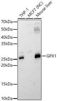
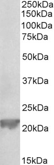
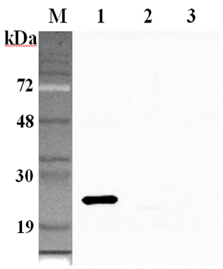
WB analysis of various samples using GTX04981 GPX1 antibody. NC: negative control Loading : 25μg Dilution : 1:1000


WB analysis of various samples using GTX04981 GPX1 antibody. NC: negative control Loading : 25μg Dilution : 1:1000
GPX1 antibody
GTX04981
ApplicationsImmunoFluorescence, Western Blot, ImmunoCytoChemistry, ImmunoHistoChemistry, ImmunoHistoChemistry Paraffin
Product group Antibodies
ReactivityHuman, Mouse, Rat
TargetGPX1
Overview
- SupplierGeneTex
- Product NameGPX1 antibody
- Delivery Days Customer7
- Application Supplier NoteWB: 1:500-1:1000. ICC/IF: 1:50-1:200. IHC-P: 1:50-1:200. *Optimal dilutions/concentrations should be determined by the researcher.Not tested in other applications.
- ApplicationsImmunoFluorescence, Western Blot, ImmunoCytoChemistry, ImmunoHistoChemistry, ImmunoHistoChemistry Paraffin
- CertificationResearch Use Only
- ClonalityPolyclonal
- ConjugateUnconjugated
- Gene ID2876
- Target nameGPX1
- Target descriptionglutathione peroxidase 1
- Target synonymsGPXD, GSHPX1, glutathione peroxidase 1, GSHPx-1, cellular glutathione peroxidase, phospholipid-hydroperoxide glutathione peroxidase GPX1, selenoprotein GPX1
- HostRabbit
- IsotypeIgG
- Protein IDP07203
- Protein NameGlutathione peroxidase 1
- Scientific DescriptionThe protein encoded by this gene belongs to the glutathione peroxidase family, members of which catalyze the reduction of organic hydroperoxides and hydrogen peroxide (H2O2) by glutathione, and thereby protect cells against oxidative damage. Other studies indicate that H2O2 is also essential for growth-factor mediated signal transduction, mitochondrial function, and maintenance of thiol redox-balance; therefore, by limiting H2O2 accumulation, glutathione peroxidases are also involved in modulating these processes. Several isozymes of this gene family exist in vertebrates, which vary in cellular location and substrate specificity. This isozyme is the most abundant, is ubiquitously expressed and localized in the cytoplasm, and whose preferred substrate is hydrogen peroxide. It is also a selenoprotein, containing the rare amino acid selenocysteine (Sec) at its active site. Sec is encoded by the UGA codon, which normally signals translation termination. The 3 UTRs of selenoprotein mRNAs contain a conserved stem-loop structure, designated the Sec insertion sequence (SECIS) element, that is necessary for the recognition of UGA as a Sec codon, rather than as a stop signal. This gene contains an in-frame GCG trinucleotide repeat in the coding region, and three alleles with 4, 5 or 6 repeats have been found in the human population. The allele with 4 GCG repeats has been significantly associated with breast cancer risk in premenopausal women. Alternatively spliced transcript variants have been found for this gene. Pseudogenes of this locus have been identified on chromosomes X and 21. [provided by RefSeq, Aug 2017]
- ReactivityHuman, Mouse, Rat
- Storage Instruction-20°C or -80°C,2°C to 8°C
- UNSPSC41116161
Datasheet
Related products
Product group Antibodies
ApplicationsWestern Blot, ELISA
ReactivityMouse, Rat
- SizePrice
Product group Antibodies
anti-GPX1 (human), pAbAG-25A-0104
ApplicationsWestern Blot, ELISA
ReactivityHuman
TargetGPX1
- SizePrice

Product group Antibodies
Anti-GPX1 Antibody Picoband(r)A01019-2-CARRIER-FREE
ApplicationsWestern Blot, ELISA, ImmunoHistoChemistry
ReactivityHuman, Mouse, Rat
TargetGPX1
- SizePrice
Product group Antibodies
Anti-GPX1 Antibody144-62010
ApplicationsWestern Blot, ImmunoHistoChemistry
ReactivityHuman, Mouse, Rat
TargetGPX1
- SizePrice

Product group Antibodies
GPX1 Recombinant AntibodyBSM-52772R
ApplicationsWestern Blot
ReactivityHuman, Mouse, Rat
TargetGPX1
- SizePrice
Product group Antibodies
GPX1 Recombinant Monoclonal AntibodyCSB-RA009866A0HU
ApplicationsELISA
ReactivityHuman
TargetGPX1
- SizePrice
Product group Antibodies

ApplicationsWestern Blot, ELISA, ImmunoHistoChemistry
ReactivityHuman, Porcine
TargetGPX1
- SizePrice
Product group Antibodies
ApplicationsImmunoPrecipitation, Western Blot, ImmunoCytoChemistry, ImmunoHistoChemistry
ReactivityMouse, Porcine, Rat
TargetGPX1
- SizePrice
Product group Antibodies

GPX1 / Glutathione Peroxidase AntibodyLS-C331264
ApplicationsWestern Blot, ImmunoHistoChemistry
ReactivityHuman, Mouse, Rat
TargetGPX1
- SizePrice
Product group Antibodies
TargetGPX1
- SizePrice