

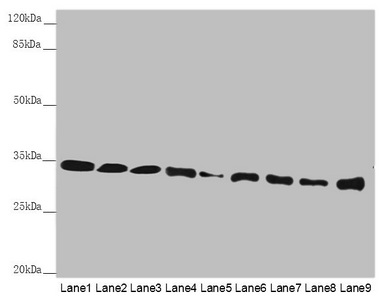
Western blot All lanes: HADH antibody at 2.38microg/ml Lane 1: Mouse heart tissue Lane 2: Mouse liver tissue Lane 3: Mouse kidney tissue Lane 4: Mouse skeletal muscle tissue Lane 5: NIH/3T3 whole cell lysate Lane 6: Hela whole cell lysate Lane 7: 293T whole cell lysate Lane 8: A431 whole cell lysate Lane 9: Jurkat whole cell lysate Secondary Goat polyclonal to rabbit IgG at 1/10000 dilution Predicted band size: 35, 43, 37 kDa Observed band size: 35 kDa


Western blot All lanes: HADH antibody at 2.38microg/ml Lane 1: Mouse heart tissue Lane 2: Mouse liver tissue Lane 3: Mouse kidney tissue Lane 4: Mouse skeletal muscle tissue Lane 5: NIH/3T3 whole cell lysate Lane 6: Hela whole cell lysate Lane 7: 293T whole cell lysate Lane 8: A431 whole cell lysate Lane 9: Jurkat whole cell lysate Secondary Goat polyclonal to rabbit IgG at 1/10000 dilution Predicted band size: 35, 43, 37 kDa Observed band size: 35 kDa
HADH Antibody
CSB-PA614992ESR1HU
ApplicationsWestern Blot, ELISA, ImmunoHistoChemistry
Product group Antibodies
ReactivityHuman, Mouse
TargetHADH
Overview
- SupplierCusabio
- Product NameHADH Antibody
- Delivery Days Customer20
- ApplicationsWestern Blot, ELISA, ImmunoHistoChemistry
- CertificationResearch Use Only
- ClonalityPolyclonal
- ConjugateUnconjugated
- Gene ID3033
- Target nameHADH
- Target descriptionhydroxyacyl-CoA dehydrogenase
- Target synonymsHAD, HADH1, HADHSC, HCDH, HHF4, MSCHAD, SCHAD, hydroxyacyl-coenzyme A dehydrogenase, mitochondrial, L-3-hydroxyacyl-Coenzyme A dehydrogenase, short chain, medium and short-chain L-3-hydroxyacyl-coenzyme A dehydrogenase, short-chain 3-hydroxyacyl-CoA dehydrogenase, testis secretory sperm-binding protein Li 203a
- HostRabbit
- IsotypeIgG
- Protein IDQ16836
- Protein NameHydroxyacyl-coenzyme A dehydrogenase, mitochondrial
- Scientific DescriptionPlays an essential role in the mitochondrial beta-oxidation of short chain fatty acids. Exerts it highest activity toward 3-hydroxybutyryl-CoA.
- ReactivityHuman, Mouse
- Storage Instruction-20°C or -80°C
- UNSPSC41116161
Related products
Product group Antibodies
Anti-HADH AntibodyA116629
ApplicationsDot Blot, ImmunoFluorescence, Western Blot, ELISA
ReactivityPorcine
- SizePrice

Product group Antibodies
Anti-HADH Antibody Picoband(r)A03650-1-CARRIER-FREE
ApplicationsFlow Cytometry, ImmunoFluorescence, Western Blot, ELISA, ImmunoCytoChemistry, ImmunoHistoChemistry
ReactivityHuman, Mouse, Rat
TargetHADH
- SizePrice
Product group Antibodies
Anti-HADH Antibody144-01076
ApplicationsWestern Blot, ImmunoHistoChemistry
ReactivityHuman, Mouse, Rat
TargetHADH
- SizePrice
Product group Antibodies
HADHSC Polyclonal AntibodyBS-10020R
ApplicationsImmunoFluorescence, Western Blot, ELISA, ImmunoCytoChemistry, ImmunoHistoChemistry, ImmunoHistoChemistry Frozen, ImmunoHistoChemistry Paraffin
ReactivityBovine, Human, Mouse, Porcine, Rat
TargetHADH
- SizePrice
Product group Antibodies

Goat anti-HADH / HADHSCEB08200
ApplicationsWestern Blot, ELISA
ReactivityCanine, Human, Mouse, Rat
TargetHADH
- SizePrice
Product group Antibodies
ApplicationsImmunoPrecipitation, Western Blot, ImmunoCytoChemistry, ImmunoHistoChemistry
ReactivityPorcine
TargetHADH
- SizePrice
Product group Antibodies

HADH AntibodyLS-C331243
ApplicationsWestern Blot, ImmunoHistoChemistry
ReactivityHuman, Mouse, Rat
TargetHADH
- SizePrice

Product group Antibodies
Anti-HADH AntibodyHPA039588
ApplicationsWestern Blot, ImmunoHistoChemistry
ReactivityHuman
TargetHADH
- SizePrice
Product group Antibodies
HADH antibodyGTX105167
ApplicationsImmunoFluorescence, ImmunoPrecipitation, Western Blot, ImmunoCytoChemistry, ImmunoHistoChemistry, ImmunoHistoChemistry Paraffin
ReactivityHuman, Mouse, Rat
TargetHADH
- SizePrice
Product group Antibodies
Anti-LAPTM4B AntibodyCAB10761
ApplicationsWestern Blot, ELISA
ReactivityHuman
TargetHADH
- SizePrice