

HAGH Antibody Pair
CSB-EAP11195
ApplicationsELISA
Product group Antibodies
ReactivityBovine, Canine, Human, Porcine
TargetHAGH
Overview
- SupplierCusabio
- Product NameHAGH Antibody Pair
- Delivery Days Customer9
- Application Supplier NoteWe recommend using the capture antibody at a concentration of 0.5ug/ml and the detection antibody at a concentration of 0.125ug/ml.Optimal dilutions should be determined experimentally by the researcher.
- ApplicationsELISA
- CertificationResearch Use Only
- ConjugateBiotin
- Gene ID3029
- Target nameHAGH
- Target descriptionhydroxyacylglutathione hydrolase
- Target synonymsGLO2, GLO2D, GLX2, GLXII, HAGH1, hydroxyacylglutathione hydrolase, mitochondrial, glyoxalase II, hydroxyacylglutathione hydroxylase
- HostRabbit
- IsotypeIgG
- Protein IDQ16775
- Protein NameHydroxyacylglutathione hydrolase, mitochondrial
- ReactivityBovine, Canine, Human, Porcine
- UNSPSC41116163
Related products
Product group Antibodies
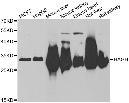
Anti-HAGH AntibodyA31462
ApplicationsWestern Blot, ImmunoHistoChemistry
ReactivityHuman, Mouse, Rat
- SizePrice

Product group Antibodies
Anti-HAGH Antibody Picoband(r)A08943-1-CARRIER-FREE
ApplicationsFlow Cytometry, Western Blot, ELISA, ImmunoHistoChemistry
ReactivityHuman, Mouse, Rat
TargetHAGH
- SizePrice
Product group Antibodies
Anti-HAGH Antibody144-06615
ApplicationsImmunoFluorescence, Western Blot, ImmunoHistoChemistry
ReactivityHuman, Mouse, Rat
TargetHAGH
- SizePrice
Product group Antibodies
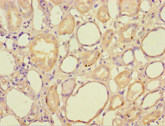
HAGH Polyclonal AntibodyBS-15404R
ApplicationsImmunoFluorescence, ELISA, ImmunoCytoChemistry, ImmunoHistoChemistry, ImmunoHistoChemistry Frozen, ImmunoHistoChemistry Paraffin
ReactivityCanine, Equine, Human, Mouse, Rat
- SizePrice
Product group Antibodies
HAGH AntibodyCSB-PA11195A0RB
ApplicationsELISA, ImmunoHistoChemistry
ReactivityHuman
TargetHAGH
- SizePrice
Product group Antibodies

HAGH AntibodyLS-C334831
ApplicationsImmunoFluorescence, Western Blot, ImmunoHistoChemistry
ReactivityHuman
TargetHAGH
- SizePrice

Product group Antibodies
Anti-HAGH AntibodyHPA043041
ApplicationsImmunoHistoChemistry
ReactivityHuman
TargetHAGH
- SizePrice
Product group Antibodies
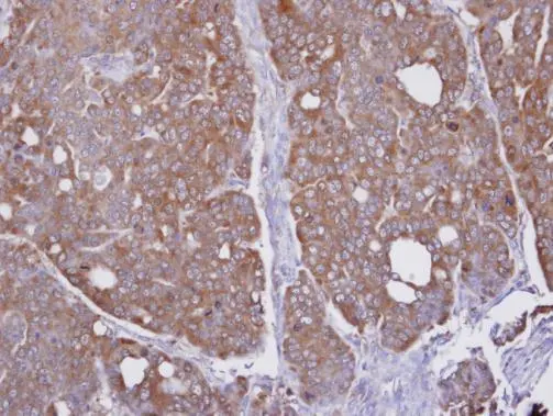
HAGH antibody [N2C3]GTX105708
ApplicationsWestern Blot, ImmunoHistoChemistry, ImmunoHistoChemistry Paraffin
ReactivityHuman, Mouse
TargetHAGH
- SizePrice
Product group Antibodies
Anti-HAGH AntibodyCAB6615
ApplicationsImmunoFluorescence, Western Blot, ELISA, ImmunoCytoChemistry
ReactivityHuman
TargetHAGH
- SizePrice