

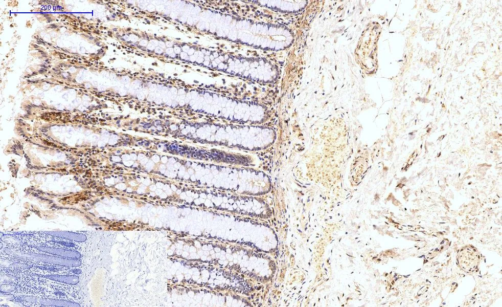
IHC-P analysis of human colon tissue using GTX33987 HAO1 antibody [Mix]. Negative control (the lower left coner) was secondary antibody only. Antigen retrieval : Sodium citrate pH6.0 was used for antibody retrieval (>98oC, 20min) Dilution : 1:200
![IHC-P analysis of rat kidney tissue using GTX33987 HAO1 antibody [Mix]. Negative control (the lower left coner) was secondary antibody only. Antigen retrieval : Sodium citrate pH6.0 was used for antibody retrieval (>98oC, 20min) Dilution : 1:200](https://www.genetex.com/upload/website/prouct_img/normal/GTX33987/GTX33987_20200622_IHC-P_220_w_23060801_705.webp "IHC-P analysis of rat kidney tissue using GTX33987 HAO1 antibody [Mix]. Negative control (the lower left coner) was secondary antibody only. Antigen retrieval : Sodium citrate pH6.0 was used for antibody retrieval (>98oC, 20min) Dilution : 1:200")
![IHC-P analysis of rat liver tissue using GTX33987 HAO1 antibody [Mix]. Negative control (the lower left coner) was secondary antibody only. Antigen retrieval : Sodium citrate pH6.0 was used for antibody retrieval (>98oC, 20min) Dilution : 1:200](https://www.genetex.com/upload/website/prouct_img/normal/GTX33987/GTX33987_20200622_IHC-P_228_w_23060801_921.webp "IHC-P analysis of rat liver tissue using GTX33987 HAO1 antibody [Mix]. Negative control (the lower left coner) was secondary antibody only. Antigen retrieval : Sodium citrate pH6.0 was used for antibody retrieval (>98oC, 20min) Dilution : 1:200")
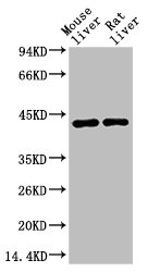
![WB analysis of mouse and rat liver tissue lysates using GTX33987 HAO1 antibody [Mix].](https://www.genetex.com/upload/website/prouct_img/normal/GTX33987/GTX33987_20200622_WB_544_w_23060801_140.webp "WB analysis of mouse and rat liver tissue lysates using GTX33987 HAO1 antibody [Mix].")
![IHC-P analysis of human uterus cancer tissue using GTX33987 HAO1 antibody [Mix]. Negative control (the lower left coner) was secondary antibody only. Antigen retrieval : Sodium citrate pH6.0 was used for antibody retrieval (>98oC, 20min) Dilution : 1:200](https://www.genetex.com/upload/website/prouct_img/normal/GTX33987/GTX33987_20200622_IHC-P_052_w_23060801_162.webp "IHC-P analysis of human uterus cancer tissue using GTX33987 HAO1 antibody [Mix]. Negative control (the lower left coner) was secondary antibody only. Antigen retrieval : Sodium citrate pH6.0 was used for antibody retrieval (>98oC, 20min) Dilution : 1:200")
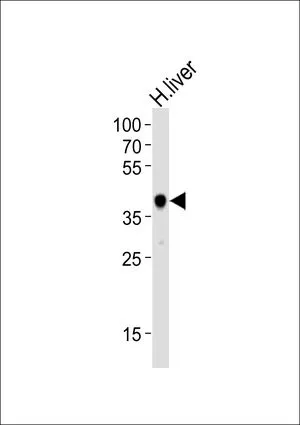
![IHC-P analysis of human liver tissue using GTX33987 HAO1 antibody [Mix]. Negative control (the lower left coner) was secondary antibody only. Antigen retrieval : Sodium citrate pH6.0 was used for antibody retrieval (>98oC, 20min) Dilution : 1:200](https://www.genetex.com/upload/website/prouct_img/normal/GTX33987/GTX33987_20200622_IHC-P_018_w_23060801_802.webp "IHC-P analysis of human liver tissue using GTX33987 HAO1 antibody [Mix]. Negative control (the lower left coner) was secondary antibody only. Antigen retrieval : Sodium citrate pH6.0 was used for antibody retrieval (>98oC, 20min) Dilution : 1:200")
![IHC-P analysis of mouse heart tissue using GTX33987 HAO1 antibody [Mix]. Negative control (the lower left coner) was secondary antibody only. Antigen retrieval : Sodium citrate pH6.0 was used for antibody retrieval (>98oC, 20min) Dilution : 1:200](https://www.genetex.com/upload/website/prouct_img/normal/GTX33987/GTX33987_20200622_IHC-P_090_w_23060801_585.webp "IHC-P analysis of mouse heart tissue using GTX33987 HAO1 antibody [Mix]. Negative control (the lower left coner) was secondary antibody only. Antigen retrieval : Sodium citrate pH6.0 was used for antibody retrieval (>98oC, 20min) Dilution : 1:200")
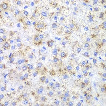
![IHC-P analysis of mouse liver tissue using GTX33987 HAO1 antibody [Mix]. Negative control (the lower left coner) was secondary antibody only. Antigen retrieval : Sodium citrate pH6.0 was used for antibody retrieval (>98oC, 20min) Dilution : 1:200](https://www.genetex.com/upload/website/prouct_img/normal/GTX33987/GTX33987_20200622_IHC-P_104_w_23060801_653.webp "IHC-P analysis of mouse liver tissue using GTX33987 HAO1 antibody [Mix]. Negative control (the lower left coner) was secondary antibody only. Antigen retrieval : Sodium citrate pH6.0 was used for antibody retrieval (>98oC, 20min) Dilution : 1:200")
IHC-P analysis of human colon tissue using GTX33987 HAO1 antibody [Mix]. Negative control (the lower left coner) was secondary antibody only. Antigen retrieval : Sodium citrate pH6.0 was used for antibody retrieval (>98oC, 20min) Dilution : 1:200
HAO1 antibody [Mix]
GTX33987
ApplicationsWestern Blot, ImmunoHistoChemistry, ImmunoHistoChemistry Paraffin
Product group Antibodies
ReactivityHuman, Mouse, Rat
TargetHAO1
Overview
- SupplierGeneTex
- Product NameHAO1 antibody [Mix]
- Delivery Days Customer9
- Application Supplier NoteWB: 1:1000-1:2000. *Optimal dilutions/concentrations should be determined by the researcher.Not tested in other applications.
- ApplicationsWestern Blot, ImmunoHistoChemistry, ImmunoHistoChemistry Paraffin
- CertificationResearch Use Only
- ClonalityPolyclonal
- Clone IDMix
- ConjugateUnconjugated
- Gene ID54363
- Target nameHAO1
- Target descriptionhydroxyacid oxidase 1
- Target synonymsGO, GOX, GOX1, HAOX1, 2-Hydroxyacid oxidase 1, (S)-2-hydroxy-acid oxidase, glycolate oxidase 1, glyoxylate oxidase, hydroxyacid oxidase (glycolate oxidase) 1
- HostMouse
- IsotypeIgG1
- Protein IDQ9UJM8
- Protein Name2-Hydroxyacid oxidase 1
- Scientific DescriptionThis gene is one of three related genes that have 2-hydroxyacid oxidase activity yet differ in encoded protein amino acid sequence, tissue expression and substrate preference. Subcellular location of the encoded protein is the peroxisome. Specifically, this gene is expressed primarily in liver and pancreas and the encoded protein is most active on glycolate, a two-carbon substrate. The protein is also active on 2-hydroxy fatty acids. The transcript detected at high levels in pancreas may represent an alternatively spliced form or the use of a multiple near-consensus upstream polyadenylation site. [provided by RefSeq, Jul 2008]
- ReactivityHuman, Mouse, Rat
- Storage Instruction-20°C or -80°C,2°C to 8°C
- UNSPSC41116161
Datasheet
Related products

Product group Antibodies
Anti-HAO1 Antibody Picoband(r)A09159-2-CARRIER-FREE
ApplicationsFlow Cytometry, Western Blot, ELISA, ImmunoHistoChemistry
ReactivityHuman, Mouse, Rat
TargetHAO1
- SizePrice
Product group Antibodies
Anti-HAO1 AntibodyA41277
ApplicationsWestern Blot
ReactivityMouse, Rat
- SizePrice
Product group Antibodies

HAO1 Antibody (clone 3B2)LS-C767091
ApplicationsImmunoFluorescence, Western Blot, ImmunoHistoChemistry, ImmunoHistoChemistry Paraffin
ReactivityMouse, Rat
TargetHAO1
- SizePrice

Product group Antibodies
Anti-HAO1 AntibodyHPA049552
ApplicationsWestern Blot, ImmunoHistoChemistry
ReactivityHuman
TargetHAO1
- SizePrice
Product group Antibodies
HAO1 Monoclonal AntibodyCSB-MA080172
ApplicationsWestern Blot, ELISA
ReactivityMouse, Rat
TargetHAO1
- SizePrice
Product group Antibodies
HAO1 antibody, InternalGTX81144
ApplicationsWestern Blot
ReactivityHuman, Mouse
TargetHAO1
- SizePrice
![WB analysis of various cell lines using GTX84392 HAO1 antibody [3E7]. Loading : 35 ug per lane](https://www.genetex.com/upload/website/prouct_img/normal/GTX84392/GTX84392_3526_WB_w_23061420_975.webp)
Product group Antibodies
HAO1 antibody [3E7]GTX84392
ApplicationsFlow Cytometry, Western Blot
ReactivityCanine, Human, Monkey, Rat
TargetHAO1
- SizePrice
Product group Antibodies
HAO1 antibodyGTX54164
ApplicationsWestern Blot, ImmunoHistoChemistry, ImmunoHistoChemistry Paraffin
ReactivityHuman, Mouse, Rat
TargetHAO1
- SizePrice