

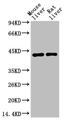
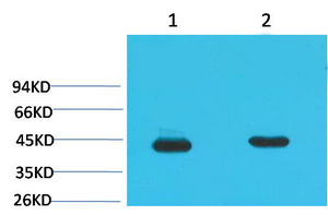
Western blot analysis of 1) Mouse Liver Tissue, 2) Rat Liver Tissue using HAO1 Monoclonal Antibody.
Western blot analysis of 1) Mouse Liver Tissue, 2) Rat Liver Tissue using HAO1 Monoclonal Antibody.
HAO1 Monoclonal Antibody
CSB-MA080172
ApplicationsWestern Blot, ELISA
Product group Antibodies
ReactivityMouse, Rat
TargetHAO1
Overview
- SupplierCusabio
- Product NameHAO1 Monoclonal Antibody
- Delivery Days Customer20
- ApplicationsWestern Blot, ELISA
- CertificationResearch Use Only
- ClonalityMonoclonal
- ConjugateUnconjugated
- Gene ID54363
- Target nameHAO1
- Target descriptionhydroxyacid oxidase 1
- Target synonymsGO, GOX, GOX1, HAOX1, 2-Hydroxyacid oxidase 1, (S)-2-hydroxy-acid oxidase, glycolate oxidase 1, glyoxylate oxidase, hydroxyacid oxidase (glycolate oxidase) 1
- HostMouse
- IsotypeIgG
- Protein IDQ9UJM8
- Protein Name2-Hydroxyacid oxidase 1
- ReactivityMouse, Rat
- Storage Instruction-20°C or -80°C
- UNSPSC41116161
Related products

Product group Antibodies
Anti-HAO1 Antibody Picoband(r)A09159-2-CARRIER-FREE
ApplicationsFlow Cytometry, Western Blot, ELISA, ImmunoHistoChemistry
ReactivityHuman, Mouse, Rat
TargetHAO1
- SizePrice
Product group Antibodies
Anti-HAO1 AntibodyA41277
ApplicationsWestern Blot
ReactivityMouse, Rat
- SizePrice
Product group Antibodies

HAO1 Antibody (clone 3B2)LS-C767091
ApplicationsImmunoFluorescence, Western Blot, ImmunoHistoChemistry, ImmunoHistoChemistry Paraffin
ReactivityMouse, Rat
TargetHAO1
- SizePrice

Product group Antibodies
Anti-HAO1 AntibodyHPA049552
ApplicationsWestern Blot, ImmunoHistoChemistry
ReactivityHuman
TargetHAO1
- SizePrice
![IHC-P analysis of human colon tissue using GTX33987 HAO1 antibody [Mix]. Negative control (the lower left coner) was secondary antibody only. Antigen retrieval : Sodium citrate pH6.0 was used for antibody retrieval (>98oC, 20min) Dilution : 1:200](https://www.genetex.com/upload/website/prouct_img/normal/GTX33987/GTX33987_20200622_IHC-P_064_w_23060801_325.webp)
Product group Antibodies
HAO1 antibody [Mix]GTX33987
ApplicationsWestern Blot, ImmunoHistoChemistry, ImmunoHistoChemistry Paraffin
ReactivityHuman, Mouse, Rat
TargetHAO1
- SizePrice
Product group Antibodies
Anti-HAO1 Antibody144-06470
ApplicationsImmunoFluorescence, Western Blot, ImmunoHistoChemistry
ReactivityHuman, Mouse, Rat
TargetHAO1
- SizePrice
Product group Antibodies
HAO1 Polyclonal AntibodyBS-8602R
ApplicationsImmunoFluorescence, Western Blot, ELISA, ImmunoCytoChemistry, ImmunoHistoChemistry, ImmunoHistoChemistry Frozen, ImmunoHistoChemistry Paraffin
ReactivityCanine, Equine, Human, Mouse, Porcine, Rabbit, Rat
- SizePrice