

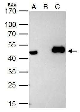
HAT1 antibody immunoprecipitates HAT1 protein in IP experiments. IP Sample: HeLa whole cell lysate/extract A. 40 μg HeLa whole cell lysate/extract B. Control with 2 μg of preimmune rabbit IgG C. Immunoprecipitation of HAT1 protein by 2 μg of HAT1 antibody (GTX110643) 12% SDS-PAGE The immunoprecipitated HAT1 protein was detected by HAT1 antibody (GTX110643) diluted at 1:1000. EasyBlot anti-rabbit IgG (GTX221666-01) was used as a secondary reagent.
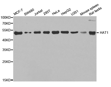
 were separated by 10% SDS-PAGE, and the membrane was blotted with HAT1 antibody (GTX110643) diluted by 1:10000.")
![HAT1 antibody [N1C3-2] detects HAT1 protein at cytoplasm and nucleus by immunofluorescent analysis. Sample: HeLa cells were fixed in 4% paraformaldehyde at RT for 15 min. Green: HAT1 protein stained by HAT1 antibody [N1C3-2] (GTX110643) diluted at 1:500. Blue: Hoechst 33342 staining.](https://www.genetex.com/upload/website/prouct_img/normal/GTX110643/GTX110643_40450_IFA_w_23060500_505.webp "HAT1 antibody [N1C3-2] detects HAT1 protein at cytoplasm and nucleus by immunofluorescent analysis. Sample: HeLa cells were fixed in 4% paraformaldehyde at RT for 15 min. Green: HAT1 protein stained by HAT1 antibody [N1C3-2] (GTX110643) diluted at 1:500. Blue: Hoechst 33342 staining.")
 antibody at 1:250 dilution.
Antigen Retrieval: Trilogy? (EDTA based, pH 8.0) buffer, 15min")
HAT1 antibody immunoprecipitates HAT1 protein in IP experiments. IP Sample: HeLa whole cell lysate/extract A. 40 μg HeLa whole cell lysate/extract B. Control with 2 μg of preimmune rabbit IgG C. Immunoprecipitation of HAT1 protein by 2 μg of HAT1 antibody (GTX110643) 12% SDS-PAGE The immunoprecipitated HAT1 protein was detected by HAT1 antibody (GTX110643) diluted at 1:1000. EasyBlot anti-rabbit IgG (GTX221666-01) was used as a secondary reagent.
HAT1 antibody
GTX110643
ApplicationsImmunoFluorescence, ImmunoPrecipitation, Western Blot, ChIP Chromatin ImmunoPrecipitation, ImmunoCytoChemistry, ImmunoHistoChemistry, ImmunoHistoChemistry Paraffin
Product group Antibodies
ReactivityHuman
TargetHAT1
Overview
- SupplierGeneTex
- Product NameHAT1 antibody
- Delivery Days Customer9
- Application Supplier NoteWB: 1:5000-1:20000. ICC/IF: 1:100-1:1000. IHC-P: 1:100-1:1000. IP: 1:100-1:500. *Optimal dilutions/concentrations should be determined by the researcher.Not tested in other applications.
- ApplicationsImmunoFluorescence, ImmunoPrecipitation, Western Blot, ChIP Chromatin ImmunoPrecipitation, ImmunoCytoChemistry, ImmunoHistoChemistry, ImmunoHistoChemistry Paraffin
- CertificationResearch Use Only
- ClonalityPolyclonal
- Concentration0.98 mg/ml
- ConjugateUnconjugated
- Gene ID8520
- Target nameHAT1
- Target descriptionhistone acetyltransferase 1
- Target synonymsKAT1, histone acetyltransferase type B catalytic subunit
- HostRabbit
- IsotypeIgG
- Protein IDO14929
- Protein NameHistone acetyltransferase type B catalytic subunit
- Scientific DescriptionHistone acetylation, particularly of histone H4, plays an important role in replication-dependent chromatin assembly. This gene encodes a histone acetylase that contains A, B, and D motifs, present in many N-acetyltransferases, including those that acetylate substrates other than histones. Alternatively spliced transcript variants that encode different isoforms have been identified for this gene. [provided by RefSeq]
- ReactivityHuman
- Storage Instruction-20°C or -80°C,2°C to 8°C
- UNSPSC41116161
Datasheet
Related products
Product group Antibodies
Anti-HAT1 AntibodyA31141
ApplicationsImmunoFluorescence, ImmunoPrecipitation, Western Blot, ChIP Chromatin ImmunoPrecipitation, ImmunoHistoChemistry
ReactivityHuman, Mouse, Rat
- SizePrice
Product group Antibodies
Anti-KAT1 (HAT1) (N-term) Antibody102-20629
ApplicationsWestern Blot
TargetHAT1
- SizePrice

Product group Antibodies
Anti-KAT1/HAT1 Antibody Picoband(r)A03596-2-CARRIER-FREE
ApplicationsFlow Cytometry, ImmunoFluorescence, Western Blot, ImmunoCytoChemistry, ImmunoHistoChemistry
ReactivityHuman, Mouse, Rat
TargetHAT1
- SizePrice
Product group Antibodies
KAT1 Recombinant Antibody, AbBy Fluor-405 ConjugatedBSM-62022R-BF405
ApplicationsFlow Cytometry, ImmunoFluorescence, Western Blot
ReactivityHuman, Mouse, Rat
TargetHAT1
- SizePrice
Product group Antibodies
HAT1 AntibodyCSB-PA010143EA01HU
ApplicationsImmunoFluorescence, Western Blot, ELISA
ReactivityHuman, Rat
TargetHAT1
- SizePrice
Product group Antibodies
HAT1 Polyclonal AntibodyCAC14843
ApplicationsImmunoFluorescence, Western Blot, ELISA
ReactivityRat
TargetHAT1
- SizePrice

Product group Antibodies
Anti-HAT1 AntibodyHPA036788
ApplicationsWestern Blot, ImmunoCytoChemistry, ImmunoHistoChemistry
ReactivityHuman, Mouse, Rat
TargetHAT1
- SizePrice
Product group Antibodies
ApplicationsImmunoFluorescence, Western Blot, ELISA, ImmunoCytoChemistry, ImmunoHistoChemistry, ImmunoHistoChemistry Paraffin
ReactivityHuman
TargetHAT1
- SizePrice
Product group Antibodies

HAT1 Antibody (aa245-419)LS-C334546
ApplicationsImmunoFluorescence, ImmunoPrecipitation, Western Blot, ChIP Chromatin ImmunoPrecipitation, ImmunoHistoChemistry
ReactivityHuman, Mouse, Rat
TargetHAT1
- SizePrice