

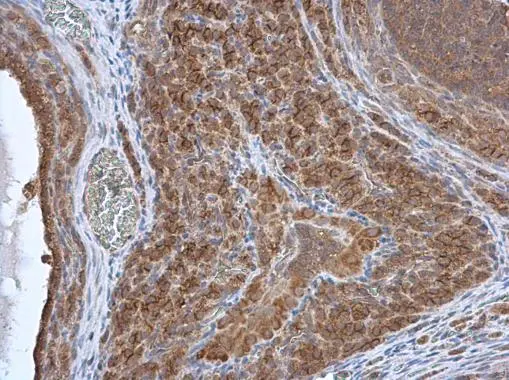
HAX1 antibody detects HAX1 protein at cytoplasm in rat ovary by immunohistochemical analysis. Sample: Paraffin-embedded rat ovary. HAX1 antibody (GTX101992) diluted at 1:500.
Antigen Retrieval: Citrate buffer, pH 6.0, 15 min
 antibody at 1:100 dilution.
Antigen Retrieval: Citrate buffer, pH 6.0, 15 min")
![HAX1 antibody detects HAX1 protein at cell membrane by immunofluorescent analysis. Sample: HeLa cells were fixed in ice-cold MeOH for 5 min. Green: HAX1 stained by HAX1 antibody (GTX101992) diluted at 1:500. Red: alpha Tubulin, a cytoskeleton marker, stained by alpha Tubulin antibody [GT114] (GTX628802) diluted at 1:1000. Blue: Fluoroshield with DAPI (GTX30920).](https://www.genetex.com/upload/website/prouct_img/normal/GTX101992/GTX101992_44454_20220121_ICC_IF_w_23060100_485.webp "HAX1 antibody detects HAX1 protein at cell membrane by immunofluorescent analysis. Sample: HeLa cells were fixed in ice-cold MeOH for 5 min. Green: HAX1 stained by HAX1 antibody (GTX101992) diluted at 1:500. Red: alpha Tubulin, a cytoskeleton marker, stained by alpha Tubulin antibody [GT114] (GTX628802) diluted at 1:1000. Blue: Fluoroshield with DAPI (GTX30920).")
 of methanol-fixed A431, using HAX1(GTX101992) antibody (Green) at 1:500 dilution. Alpha-tubulin filaments were labeled with GTX11304 (Red) at 1:2000.")
 diluted at 1:500.")
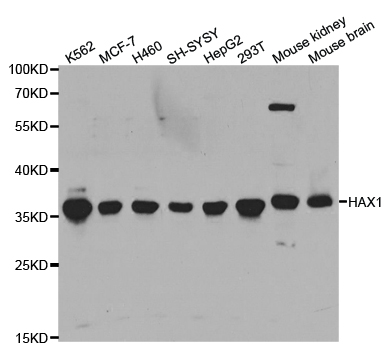
 were separated by 12% SDS-PAGE, and the membrane was blotted with HAX1 antibody (GTX101992) diluted at 1:1000. The HRP-conjugated anti-rabbit IgG antibody (GTX213110-01) was used to detect the primary antibody.")
HAX1 antibody detects HAX1 protein at cytoplasm in rat ovary by immunohistochemical analysis. Sample: Paraffin-embedded rat ovary. HAX1 antibody (GTX101992) diluted at 1:500.
Antigen Retrieval: Citrate buffer, pH 6.0, 15 min
HAX1 antibody
GTX101992
ApplicationsImmunoFluorescence, Western Blot, ImmunoCytoChemistry, ImmunoHistoChemistry, ImmunoHistoChemistry Paraffin
Product group Antibodies
ReactivityHuman, Mouse, Rat
TargetHAX1
Overview
- SupplierGeneTex
- Product NameHAX1 antibody
- Delivery Days Customer9
- Application Supplier NoteWB: 1:500-1:3000. ICC/IF: 1:100-1:1000. IHC-P: 1:100-1:1000. *Optimal dilutions/concentrations should be determined by the researcher.Not tested in other applications.
- ApplicationsImmunoFluorescence, Western Blot, ImmunoCytoChemistry, ImmunoHistoChemistry, ImmunoHistoChemistry Paraffin
- CertificationResearch Use Only
- ClonalityPolyclonal
- Concentration1.43 mg/ml
- ConjugateUnconjugated
- Gene ID10456
- Target nameHAX1
- Target descriptionHCLS1 associated protein X-1
- Target synonymsHCLSBP1, HS1BP1, SCN3, HCLS1-associated protein X-1, HAX-1, HCLS1 (and PKD2) associated protein, HS1 binding protein, HS1-associating protein X-1, HS1-binding protein 1, HSP1BP-1
- HostRabbit
- IsotypeIgG
- Protein IDO00165
- Protein NameHCLS1-associated protein X-1
- Scientific DescriptionThe protein encoded by this gene is known to associate with hematopoietic cell-specific Lyn substrate 1, a substrate of Src family tyrosine kinases. It also interacts with the product of the polycystic kidney disease 2 gene, mutations in which are associated with autosomal-dominant polycystic kidney disease, and with the F-actin-binding protein, cortactin. It was earlier thought that this gene product is mainly localized in the mitochondria, however, recent studies indicate it to be localized in the cell body. Mutations in this gene result in autosomal recessive severe congenital neutropenia, also known as Kostmann disease. Two transcript variants encoding different isoforms have been found for this gene. [provided by RefSeq]
- ReactivityHuman, Mouse, Rat
- Storage Instruction-20°C or -80°C,2°C to 8°C
- UNSPSC41116161
Datasheet
Related products
Product group Antibodies
Anti-HAX1 AntibodyA30813
ApplicationsWestern Blot, ImmunoHistoChemistry
ReactivityHuman, Mouse, Rat
- SizePrice
Product group Antibodies
Anti-HAX1 Antibody144-05551
ApplicationsImmunoFluorescence, Western Blot, ImmunoHistoChemistry
ReactivityHuman, Mouse
TargetHAX1
- SizePrice
Product group Antibodies
HAX1 Polyclonal AntibodyBS-7626R
ApplicationsImmunoFluorescence, Western Blot, ELISA, ImmunoCytoChemistry, ImmunoHistoChemistry, ImmunoHistoChemistry Frozen, ImmunoHistoChemistry Paraffin
ReactivityBovine, Canine, Equine, Human, Mouse, Porcine, Rabbit, Rat
TargetHAX1
- SizePrice

Product group Antibodies
Anti-HAX1 Antibody Picoband(r)A01495-2-CARRIER-FREE
ApplicationsFlow Cytometry, Western Blot, ELISA, ImmunoHistoChemistry
ReactivityHuman
TargetHAX1
- SizePrice
Product group Antibodies

Goat anti-HAX1EB09160
ApplicationsWestern Blot, ELISA
ReactivityHuman
TargetHAX1
- SizePrice
Product group Antibodies
HAX1 Polyclonal AntibodyCAC14042
ApplicationsImmunoFluorescence, Western Blot, ELISA, ImmunoHistoChemistry
ReactivityMouse, Rat
TargetHAX1
- SizePrice
Product group Antibodies
HAX1 AntibodyCSB-PA03515A0RB
ApplicationsImmunoFluorescence, Western Blot, ELISA, ImmunoHistoChemistry
ReactivityHuman, Mouse, Rat
TargetHAX1
- SizePrice
Product group Antibodies

HAX-1 AntibodyLS-C334130
ApplicationsImmunoFluorescence, Western Blot, ImmunoHistoChemistry
ReactivityHuman, Mouse
TargetHAX1
- SizePrice
![WB analysis of human heart tissue lysate using GTX84982 Hax1a antibody [9G6C6]. Working concentration : (A) 1 and (B) 2 μg/ml](https://www.genetex.com/upload/website/prouct_img/normal/GTX84982/GTX84982_3297_WB_20180221_w_23061420_916.webp)
Product group Antibodies
Hax1a antibody [9G6C6]GTX84982
ApplicationsWestern Blot, ELISA
ReactivityHuman, Rat
TargetHAX1
- SizePrice
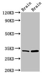
![WB analysis of human brain tissue lysate using GTX84983 Hax1a antibody [9G3D11]. Working concentration : (A) 1 and (B) 2 μg/ml](https://www.genetex.com/upload/website/prouct_img/normal/GTX84983/GTX84983_3298_WB_20180221_w_23061420_448.webp)
Product group Antibodies
Hax1a antibody [9G3D11]GTX84983
ApplicationsWestern Blot, ELISA
ReactivityHuman, Rat
TargetHAX1
- SizePrice