

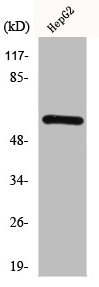
HDAC2 Antibody (Center)
MBS9231833
Product group Antibodies
Overview
- SupplierMyBioSource
- Product NameHDAC2 Antibody (Center)
- Delivery Days Customer16
- CertificationResearch Use Only
- Storage Instruction2°C to 8°C,-20°C
- UNSPSC12352203
Related products
Product group Antibodies

Goat anti-Hdac2 (mouse)EB11448
ApplicationsFlow Cytometry, ImmunoFluorescence, Western Blot, ELISA
TargetHDAC2
- SizePrice
Product group Antibodies
ApplicationsImmunoPrecipitation, Western Blot, ImmunoCytoChemistry, ImmunoHistoChemistry
TargetHDAC2
- SizePrice
Product group Antibodies
References
HDAC2 antibodyGTX109642
ApplicationsImmunoFluorescence, ImmunoPrecipitation, Western Blot, ChIP Chromatin ImmunoPrecipitation, ImmunoCytoChemistry, ImmunoHistoChemistry, ImmunoHistoChemistry Paraffin
TargetHDAC2
- SizePrice

Product group Antibodies
Anti-HDAC2 AntibodyHPA011727
ApplicationsWestern Blot, ImmunoCytoChemistry, ImmunoHistoChemistry
TargetHDAC2
- SizePrice
Product group Antibodies
HDAC2 AntibodyCSB-PA002879
ApplicationsWestern Blot, ELISA, ImmunoHistoChemistry
TargetHDAC2
- SizePrice

Product group Antibodies
Anti-HDAC2 Antibody Picoband(r)A00325-3-CARRIER-FREE
ApplicationsFlow Cytometry, ImmunoFluorescence, Western Blot, ELISA, ImmunoCytoChemistry, ImmunoHistoChemistry
TargetHDAC2
- SizePrice
Product group Antibodies
Anti-HDAC2 AntibodyA286069
ApplicationsFlow Cytometry, ImmunoFluorescence, Western Blot, ELISA
- SizePrice
Product group Antibodies
ApplicationsImmunoPrecipitation
TargetHDAC2
- SizePrice