

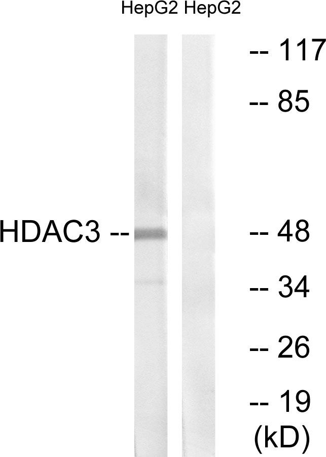
Whole cell extract (30 μg) was separated by 10% SDS-PAGE, and the membrane was blotted with HDAC3 antibody [C3], C-term (GTX109679) diluted at 1:500. The HRP-conjugated anti-rabbit IgG antibody (GTX213110-01) was used to detect the primary antibody.
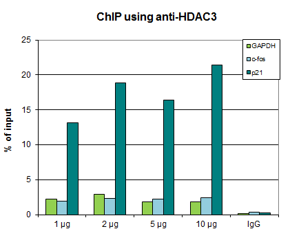
 The precipitated DNA was detected by PCR with primer set targeting to p21 promoter.")
![Various whole cell extracts (30 μg) were separated by 10% SDS-PAGE, and the membrane was blotted with HDAC3 antibody [C3], C-term (GTX109679) diluted at 1:1000. The HRP-conjugated anti-rabbit IgG antibody (GTX213110-01) was used to detect the primary antibody.](https://www.genetex.com/upload/website/prouct_img/normal/GTX109679/GTX109679_41220_20180601_WB_M_w_23060500_431.webp "Various whole cell extracts (30 μg) were separated by 10% SDS-PAGE, and the membrane was blotted with HDAC3 antibody [C3], C-term (GTX109679) diluted at 1:1000. The HRP-conjugated anti-rabbit IgG antibody (GTX213110-01) was used to detect the primary antibody.")
![HDAC3 antibody [C3], C-term detects HDAC3 protein at nucleus by immunofluorescent analysis. Sample: HeLa cells were fixed in 4% paraformaldehyde at RT for 15 min. Green: HDAC3 protein stained by HDAC3 antibody [C3], C-term (GTX109679) diluted at 1:500. Blue: Hoechst 33342 staining.](https://www.genetex.com/upload/website/prouct_img/normal/GTX109679/GTX109679_41220_IFA_w_23060500_747.webp "HDAC3 antibody [C3], C-term detects HDAC3 protein at nucleus by immunofluorescent analysis. Sample: HeLa cells were fixed in 4% paraformaldehyde at RT for 15 min. Green: HDAC3 protein stained by HDAC3 antibody [C3], C-term (GTX109679) diluted at 1:500. Blue: Hoechst 33342 staining.")
![Various whole cell extracts (30 μg) were separated by 10% SDS-PAGE, and the membrane was blotted with HDAC3 antibody [C3], C-term (GTX109679) diluted at 1:1000. The HRP-conjugated anti-rabbit IgG antibody (GTX213110-01) was used to detect the primary antibody.](https://www.genetex.com/upload/website/prouct_img/normal/GTX109679/GTX109679_39974_20180601_WB_R_w_23060500_744.webp "Various whole cell extracts (30 μg) were separated by 10% SDS-PAGE, and the membrane was blotted with HDAC3 antibody [C3], C-term (GTX109679) diluted at 1:1000. The HRP-conjugated anti-rabbit IgG antibody (GTX213110-01) was used to detect the primary antibody.")
 A: 293T B: A431 (GTX27909) C: H1299 D: HeLa S3 (GTX14654) E: HepG2 (GTX27900) F: MOLT4 (GTX27912) G: Raji (GTX27908) 10% SDS PAGE GTX109679 diluted at 1:1000 The HRP-conjugated anti-rabbit IgG antibody (GTX213110-01) was used to detect the primary antibody.")
 10% SDS-PAGE The immunoprecipitated HDAC3 protein was detected by HDAC3 antibody (GTX109679) diluted at 1:1000. EasyBlot anti-rabbit IgG (GTX221666-01) was used as a secondary reagent.")
![Non-transfected (–) and transfected (+) 293T whole cell extracts (30 μg) were separated by 10% SDS-PAGE, and the membrane was blotted with HDAC3 antibody [C3], C-term (GTX109679) diluted at 1:500. The HRP-conjugated anti-rabbit IgG antibody (GTX213110-01) was used to detect the primary antibody.](https://www.genetex.com/upload/website/prouct_img/normal/GTX109679/GTX109679_39974_20161103_WB_shRNA_watermark_w_23060500_907.webp "Non-transfected (–) and transfected (+) 293T whole cell extracts (30 μg) were separated by 10% SDS-PAGE, and the membrane was blotted with HDAC3 antibody [C3], C-term (GTX109679) diluted at 1:500. The HRP-conjugated anti-rabbit IgG antibody (GTX213110-01) was used to detect the primary antibody.")
![HDAC3 antibody [C3], C-term detects HDAC3 protein at nucleus by immunofluorescent analysis. Sample: SK-N-SH cells were fixed in 4% paraformaldehyde at RT for 15 min. Green: HDAC3 protein stained by HDAC3 antibody [C3], C-term (GTX109679) diluted at 1:400. Red: Phalloidin, a cytoskeleton marker, diluted at 1:200. Scale bar = 10 μm.](https://www.genetex.com/upload/website/prouct_img/normal/GTX109679/GTX109679_39974_20160325_IFA_w_23060500_290.webp "HDAC3 antibody [C3], C-term detects HDAC3 protein at nucleus by immunofluorescent analysis. Sample: SK-N-SH cells were fixed in 4% paraformaldehyde at RT for 15 min. Green: HDAC3 protein stained by HDAC3 antibody [C3], C-term (GTX109679) diluted at 1:400. Red: Phalloidin, a cytoskeleton marker, diluted at 1:200. Scale bar = 10 μm.")
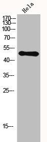
 antibody at 1:500 dilution.
Antigen Retrieval: Trilogy? (EDTA based, pH 8.0) buffer, 15min")
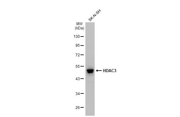
Whole cell extract (30 μg) was separated by 10% SDS-PAGE, and the membrane was blotted with HDAC3 antibody [C3], C-term (GTX109679) diluted at 1:500. The HRP-conjugated anti-rabbit IgG antibody (GTX213110-01) was used to detect the primary antibody.
HDAC3 antibody [C3], C-term
GTX109679
ApplicationsImmunoFluorescence, ImmunoPrecipitation, Western Blot, ChIP Chromatin ImmunoPrecipitation, ImmunoCytoChemistry, ImmunoHistoChemistry, ImmunoHistoChemistry Paraffin
Product group Antibodies
ReactivityDrosophila, Human, Mouse, Rat
TargetHDAC3
Overview
- SupplierGeneTex
- Product NameHDAC3 antibody [C3], C-term
- Delivery Days Customer9
- Application Supplier NoteWB: 1:500-1:3000. ICC/IF: 1:100-1:1000. IHC-P: 1:100-1:1000. IP: 1:100-1:500. *Optimal dilutions/concentrations should be determined by the researcher.Not tested in other applications.
- ApplicationsImmunoFluorescence, ImmunoPrecipitation, Western Blot, ChIP Chromatin ImmunoPrecipitation, ImmunoCytoChemistry, ImmunoHistoChemistry, ImmunoHistoChemistry Paraffin
- CertificationResearch Use Only
- ClonalityPolyclonal
- Concentration1 mg/ml
- ConjugateUnconjugated
- Gene ID8841
- Target nameHDAC3
- Target descriptionhistone deacetylase 3
- Target synonymsHD3, KDAC3, RPD3, RPD3-2, histone deacetylase 3, SMAP45, protein deacetylase HDAC3, protein deacylase HDAC3
- HostRabbit
- IsotypeIgG
- Protein IDO15379
- Protein NameHistone deacetylase 3
- Scientific DescriptionHistones play a critical role in transcriptional regulation, cell cycle progression, and developmental events. Histone acetylation/deacetylation alters chromosome structure and affects transcription factor access to DNA. The protein encoded by this gene belongs to the histone deacetylase/acuc/apha family. It has histone deacetylase activity and represses transcription when tethered to a promoter. It may participate in the regulation of transcription through its binding with the zinc-finger transcription factor YY1. This protein can also down-regulate p53 function and thus modulate cell growth and apoptosis. This gene is regarded as a potential tumor suppressor gene. [provided by RefSeq]
- ReactivityDrosophila, Human, Mouse, Rat
- Storage Instruction-20°C or -80°C,2°C to 8°C
- UNSPSC41116161
Datasheet
Related products
Product group Antibodies
Anti-HDAC3 AntibodyA95226
ApplicationsImmunoFluorescence, Western Blot, ELISA, ImmunoHistoChemistry
ReactivityHuman, Mouse, Rat
- SizePrice

Product group Antibodies
Anti-HDAC3 Antibody Picoband(r)A00839-CARRIER-FREE
ApplicationsFlow Cytometry, Western Blot, ELISA
ReactivityHuman, Mouse, Rat
TargetHDAC3
- SizePrice
Product group Antibodies
Anti-HDAC3 Antibody144-02139
ApplicationsImmunoFluorescence, ImmunoPrecipitation, Western Blot, ChIP Chromatin ImmunoPrecipitation, ImmunoHistoChemistry
ReactivityHuman, Mouse, Rat
TargetHDAC3
- SizePrice
Product group Antibodies
ApplicationsImmunoFluorescence, ChIP Chromatin ImmunoPrecipitation
ReactivityHuman
TargetHDAC3
- SizePrice
Product group Antibodies
ApplicationsFlow Cytometry, Western Blot, ImmunoCytoChemistry
ReactivityHuman, Mouse, Rat
TargetHDAC3
- SizePrice
Product group Antibodies
HDAC3 AntibodyCSB-PA002880
ApplicationsImmunoFluorescence, Western Blot, ELISA, ImmunoHistoChemistry
ReactivityHuman, Mouse, Rat
TargetHDAC3
- SizePrice
Product group Antibodies

Goat anti-HDAC3EB12818
ApplicationsWestern Blot, ELISA
ReactivityBovine, Canine, Human, Mouse, Porcine, Rat
TargetHDAC3
- SizePrice
Product group Antibodies
HDAC3 Polyclonal AntibodyCAC13761
ApplicationsImmunoFluorescence, Western Blot, ChIP Chromatin ImmunoPrecipitation, ELISA, ImmunoHistoChemistry
ReactivityMouse
TargetHDAC3
- SizePrice
![Various whole cell extracts (30 μg) were separated by 10% SDS-PAGE, and the membrane was blotted with HDAC3 antibody [GT1144] (GTX00834) diluted at 1:1000. The HRP-conjugated anti-rabbit IgG antibody (GTX213110-01) was used to detect the primary antibody, and the signal was developed with Trident ECL plus-Enhanced.](https://www.genetex.com/upload/website/prouct_img/normal/GTX00834/GTX00834_4000000016_20200410_WB_w_23053121_498.webp)
Product group Antibodies
HDAC3 antibody [GT1144]GTX00834
ApplicationsWestern Blot
ReactivityHuman, Mouse, Rat
TargetHDAC3
- SizePrice
Product group Antibodies

HDAC3 Antibody (phospho-Ser424)LS-C359014
ApplicationsImmunoFluorescence, Western Blot, ImmunoCytoChemistry, ImmunoHistoChemistry, ImmunoHistoChemistry Paraffin
ReactivityChicken, Human, Mouse, Rat
TargetHDAC3
- SizePrice